The unpaired bones of the cerebral part of the skull are. Skull: the structure of the bones of the head
The cerebral part of the skull of an adult is made up of the following bones: frontal, occipital, sphenoid, ethmoid, two temporal and two parietal. The unpaired air-bearing frontal bone consists of scales, two horizontal orbital parts and a nasal part.
The frontal bone forms the forehead and bears the frontal tubercles, which are a characteristic feature of Homo sapiens, and also forms the upper walls of the orbits, the nasal cavity, the temporal fossae, the lower and anterior walls of the anterior cranial fossa.
The paired parietal bone is involved in the formation of the roof (vault) of the skull, in the center of the bone is the parietal tubercle. The parietal bones are involved in the formation of the cranial vault.
The occipital bone, which is involved in the formation of the vault and base of the skull, the posterior cranial fossa, consists of four parts (basilar in front, two lateral and scales) located around the large (occipital) foramen. Two oval occipital condyles, located on the outer surface of the lateral parts, articulate with the atlas, forming the atlantooccipital joints, in which nodding movements and lateral tilts of the head are performed.
The unpaired air-bearing sphenoid bone has a body, on the upper surface of which there is a pituitary fossa, where the pituitary gland lies. Large wings depart from the body to the sides, upward and laterally - small wings, downward - pterygoid processes. The sphenoid bone is involved in the formation of the orbit, anterior cranial, infratemporal and pterygopalatine fossae.
The most complex is the paired air-bearing temporal bone, which is involved in the formation of the vault and base of the skull and is the seat of the organ of hearing and balance. It consists of a pyramid in which the tympanic cavity and the inner ear are located; tympanic part, in which the outer auditory opening and outdoor ear canal; squamous part, on the outer surface of which there is a deep mandibular fossa, which includes the condylar process of the lower jaw, forming the temporomandibular joint. The zygomatic process, connecting with the zygomatic bone, forms the zygomatic arch. Unpaired airway ethmoid bone consists of many cells (ethmoid labyrinths), as if suspended from the ethmoid plate, through the openings of which enter the cranial cavity olfactory nerves. From the medial surfaces of the labyrinths on both sides depart thin curved upper and middle nasal conchas, hanging down into the nasal cavity.
Some bones in the human skeleton are paired. The bones of the pelvis, upper and lower extremities, collarbones, shoulder blades. This also applies to the ribs - a person has 12 pairs of them.
There was once a belief, which some people still share today, that men have one less rib than women. The legend connects this with the biblical story of the creation of Eve from Adam's rib. Back in the 16th century, "father modern anatomy» A. Vesalius proved that this is not so: men and women have the same number of ribs.
However, sometimes people are born with the 13th unpaired rib. This anomaly is called Adam's rib syndrome, although it occurs with the same frequency in both men and women. Living with an extra rib is not easy, it compresses the nerves, blood vessels and even internal organs, interfering with their work and causing numbness of the hands. Such people undergo surgery to remove an unpaired rib.
Unpaired bones of the skeleton
Unpaired are those bones that are located in the middle of the skeleton.
Unpaired bones include all vertebrae that make up spinal column: 7 cervical vertebrae, 12 thoracic and 5 lumbar.
At the base of the spine in the form of a wedge between pelvic bones there is a large triangular bone - the sacrum. This is one of those bones by which you can determine whether the skeleton is female or male. In women, the sacrum is shorter and wider than in men, and the male sacrum is more curved than the female.
The upper part of the sacrum is connected to the last vertebra, and the lower part is connected to the coccyx, another unpaired bone. The coccyx also consists of several rudimentary vertebrae. This is the “remainder” of the tail, inherited by man from evolutionary ancestors.
Despite its rudimentary origin, the coccyx is not a useless part of the skeleton. Some muscles are attached to it, it takes on part of the load when a person sits or bends over.
Unpaired bones of the skull
The human skull is a very complex structure, consisting of many bones. Some of them are also unpaired.
The skull is divided into two sections - cerebral and facial. Unpaired bones of the facial region - occipital, frontal and sphenoid. On frontal bone special stop should be made. It does not immediately become a single whole; at the birth of a person, this bone consists of two halves connected by a seam. In this form, it is easier for the skull to pass through birth canal, less likely birth trauma. By the age of five, the seam is overgrown, and only in 10% of people it remains until the age of 40.
The unpaired bones of the facial region are the vomer that forms the lower back bony nasal septum, the hyoid bone, located under the muscle of the tongue and shaped like a horseshoe, as well as the lower jaw. This is very interesting bone. Of all the bones of the skull, only it is movably articulated with other bones, and most importantly, a number of its features “elevate” a person above other primates.
When artists want to depict a person endowed with great physical force, but not distinguished by intelligence, usually draw a person with a large, massive lower jaw. Such a sign is not accidental, it evokes an association with a monkey. Indeed, the size and mass of the lower jaw relative to the skull as a whole is much smaller in humans than in monkeys. Its small size made articulate speech possible. True, chewing food turned out to be difficult, which forced the evolutionary ancestors of man to create stone knives and master the processing of food on fire. Thus, small size the lower jaw has become one of the "engines" of human development.
Everyone will agree that the head of every person plays in his life at least important function than a heart. Actually a human skull a complex system, which has a very interesting structure and performing serious functions. The bones of the head protect the brain and sense organs. Between themselves, they are connected by seams and provide support for the digestive and respiratory systems am.
The skull is divided into the facial and brain sections. The bones of the brain part form a cavity for the brain and partly for the sense organs. In addition, they serve as the basis of the face and skeleton of the initial sections of the digestive and respiratory systems. Some cranial bones have cavities that are filled with air. They are connected to the nasal cavity. Due to this structure of the bones, the mass of the skull is not very large, but at the same time, its strength does not become less because of this. The brain skull consists of eight bones: two temporal, two parietal, frontal, sphenoid, ethmoid and occipital bones. Some bones of the facial part of the skull serve as the basis of the skeleton of the masticatory apparatus. Other bones are smaller in size and make up the cavity of the facial skull. Consider the anatomy of these two departments in more detail.
Bones of the cerebral cranial region
So, the brain section consists of eight bones:
- frontal;
- occipital;
- wedge-shaped;
- lattice;
- two temporal;
- two parietal.
Top part cerebral skull is called its vault, in another way, the roof. Bottom part is its foundation. Between the arch and the base there is a conditional line passing through the occipital external protrusion, along the nuchal upper line to the base of the mastoid process. Then the line continues above the auditory external opening, along the base of the zygomatic process and along the crest of the infratemporal view of the main wing. sphenoid bone. The line reaches the nasofrontal suture along the infraorbital margin.
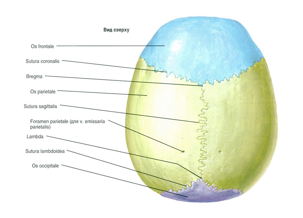
The anatomy of the cranial vault involves its division into several bones. It is half an ellipsoid in shape. Its long axis is directed to the fronto-occipital part. It corresponds to the longitudinal diameter of the brain box. Two more axes run vertically and transversely. The cranial vault has morpho-functional areas:
- unpaired fronto-parieto-occipital region;
- paired temporal region.
They are separated by temporal lines and differ in relief, mechanical conditions And bone structure. The bones of the arch have a three-layer structure. There is an inner and outer compact plate, which have a diploe between them, that is, a spongy substance. IN different areas of the arch, the ratio of compact plates and the thickness of the diploe differs. It all depends on individual variability.
It is proved that the diploe is well developed in the parasagittal zone, where the outer plate is thicker than the inner one. The lateral sections of the arch have an inverse relationship. The diploe is less in the temporal parts.
Structural features of bones determine their strength. Studies have been conducted that have proven that the compression strength of the occipital and parietal bones is greater than that of the frontal bone. The inner plate is more brittle. Even if there is no external damage, there may be comminuted fracture such a plate. This gave reason to call it a vitreous plate.
In the anatomy of the bones of the brain skull importance assigned to spongy bone. There are diploic channels. They contain diploic veins. The following important diploic canals are distinguished in the cranial vault:
- frontal;
- front;
- posterior temporal;
- occipital
Diploic channels are separated by functional feature. In this regard, it is possible to distinguish outgoing, depositing and communicating channels. They pass through suture lines in the cranial base. They are able to divide into several branches. In the outer part of the skull, the relief varies individually depending on age and sex.
The inner cranial part has a more complex relief. IN varying degrees brain elevations and finger-like impressions can be expressed. Arterial grooves, branching in a tree-like manner, originate in the cranial base from the spinous foramen. It passes through the meningeal middle artery. Dimples of granulations can be seen in the structure of the inner cranial surface. They are very changeable. In small dimples there are single growths of arachnoid meninges. In large dimples, these growths accumulate.
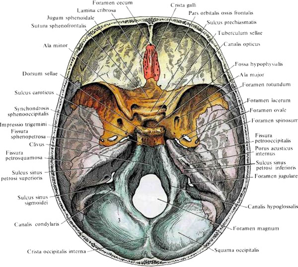
The base of the skull also has two surfaces - internal and external. The inner surface, as in the case of the cranial vault, reflects the shape of the head. It has indentations and elevations. Three pits are distinguished from the localization.
- The anterior fossa is the bed frontal lobes hemispheres of the brain. It is formed by the orbital parts of the frontal bone, part of the body of the sphenoid bone, the cribriform plate and the upper surface of the lesser wings. In the middle of the wedge-shaped protrusion, there is a border between the middle and anterior fossae.
- Middle hole. It is formed by the body of the sphenoid bone, the anterior surface of the hard stony part temporal region, small and large wings and the lower area of the scales temporal bone. In the middle fossa there are lateral and middle departments. In the lateral sections are located temporal lobes hemispheres.
- Posterior hole. It is mainly formed occipital bone. However, the body of the sphenoid bone and the petrous parts of the temporal bone type take part in this. The posterior fossa contains the cerebellum and brain stem.
There are three sections at the outer base of the skull.
- The anterior section is connected to the facial bones. It forms the nasal cavities and the roof of the eye sockets.
- Middle department. It originates at the base of the pterygoid processes and runs to a line that extends through mastoid processes, as well as the leading edge of the main hole.
- Rear section. It is formed by the temporal and occipital bones. It has three areas - mastoid, nuchal and occipital-temporal.
There are many small and large arteries at the base of the skull. Through them pass blood and cranial nerves. The thickness of the bone varies different places. The structure of stronger sections is a system of longitudinal beams converging to the body of the wedge-shaped bone. They are fastened with crossbars running transversely to the borders between the fossae of the skull. recesses cranial pits have fragile spots. It is there that fractures often occur, because the bone is quite thin. In the anterior fossa, injuries form, affecting the cribriform plate. In the middle fossa, fractures transversely pass through the back of the area, which is called the "Turkish saddle". In the posterior fossa, fractures affect the openings, and the top of the pyramid breaks off.
The Turkish saddle is located in the center of the inner base of the skull. In front, it is limited by the tubercle of the saddle. Sloping anterior processes hang over it. Behind it is limited by the back of the saddle. In the center of the saddle there is a pituitary fossa. It is the seat of the pituitary gland, that is, the endocrine gland.
Features of the cranial structure
 Of course, the structure of the entire skull is amazing, however, main feature The anatomy of the skull are pneumatic bones containing cells or air sinuses. Most of these sinuses communicate with the nasal cavity and play the role of adnexal cavities. Their role is very important - they have an aerodynamic effect on the inhaled air, so the air stream comes into contact with the olfactory receptors, which are located in the mucous membrane of the nasal cavity, more precisely, in its upper part. The paranasal sinuses are often exposed pathological processes leading to intracranial complications such as brain abscess and meningitis.
There are five main parts.
- Frontal sinus. This is a steam cavity, which is divided by a septum. Also in this part is the middle nasal passage. The sinus can be located in different places, as its length varies, - in superciliary arches, frontal scales and orbital part of the bone of the frontal type. There are single-chamber and multi-chamber sinuses.
- Sphenoid sinus. Its location is the body of the sphenoid bone. There may be additional partitions in the sinus.
- lattice cells. Their opening occurs in the middle and upper nasal passages.
- Mastoid cells. Their message from tympanic cavity occurs through the mastoid cave. Cells may vary in size. There are diploic, compact, mixed and pneumatic mastoid processes.
- Maxillary sinus. This is the largest accessory cavity of the nose.
The structure of the facial cranial region
The structure of the facial region is associated with the development of the jaws, nasal cavity, digestive and respiratory systems. The speech function also leaves an imprint on this department. Some features of the anatomy of the lower jaw are associated with the muscles that are involved in speech. The facial skull includes three main sections.
- Orbital-temporal department. These are the orbit, the anterior deepening of the temporal fossa, the anterior cranial middle fossa, the pterygopalatine and infratemporal fossae.
- Nose section. These are the paranasal sinuses, the nasal cavity and the nose itself.
- Jaw - zygomatic bones, lower and upper jaws.
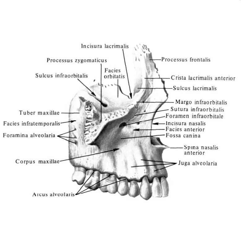 The upper jaw is an important part of the face and nasal cavity. IN different parts jaw unequal ratio of spongy and compact substance. The alveolar process has a powerful layer of spongy substance, from where it passes into the following processes. The frontal process has very small cells of spongy substance. Spongy substance from the zygomatic process goes to the infraorbital region, from where it extends almost to the frontal process. Beams of the spongy substance of the jaw are mainly located under different angles. They are grouped into the lateral and medial systems.
Lower jaw is the solid foundation of the lower facial area. She is the one in more defines the face shape. Signs of the lower jaw are a decrease in its massiveness, an increase in the angle of the branch, the presence of a chin spine, and so on. The lower jaw is the only movable part of the facial skeleton. Many muscles are attached to it, especially chewing muscles, because the configuration depends on them. The lower jaw is characterized by a basal arch. The channel in which the nerves and blood vessels pass is removed from the dental roots, but there are exceptions. The mental foramen is the exit from the jaw canal. It may be missing on one side, sometimes on both sides. On one side there may be additional holes. The ratio of spongy and compact substance is also not the same in different parts of the jaw. The outer compact plate is thicker than the inner one.
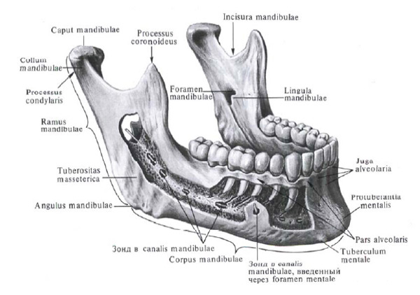
There is also a temporomandibular joint. It is formed articular surfaces head of the jaw, as well as the mandibular fossa of the temporal bone type. These surfaces are covered with fibrous cartilage. There is an articular disc, with the help of which the joint cavity is divided into the lower and upper compartments. It fuses with the joint capsule.
This is a brief excursion into the anatomy of the human skull. As we have seen, the head is a complex system consisting of different bones, joints and other elements. Everything is very interconnected, therefore, if one part of the skull suffers, this affects not only its entire condition, but also the entire body. Therefore, let's protect our heads from all kinds of injuries!


