What is fascioliasis and how to treat it? Description of fascioliasis in humans and animals
Fascioliasis is an acute and chronic zoonotic trematode disease of domestic and wild artiodactyls, accompanied by disruption of the digestive system, edema, and anemia, which reduces their productivity.
Epizootology. The source of the pathogen is animals infected with Fasciola. Among farm animals, the most susceptible to the disease are small and large horned livestock, and to a lesser extent, pigs, horses, etc. The lowest level of infestation with smallmouths occurs in the spring; by autumn it gradually increases. Scatologically, fascioliasis begins to be diagnosed in veterinary laboratories at the end of November and December. Young animals are affected significantly less than adult animals. With age, the intensity of invasion in animals increases.
The main pathophysiological, biochemical and functional changes during fascioliasis primarily occur in the liver, and only after this do disturbances occur in the activity of other organs and systems of the animal’s body.
Depending on the intensity of invasion, the resistance of the animal’s body and the stage of the disease, when conducting a blood test, we note a decrease in the number of red blood cells, hemoglobin, calcium and phosphorus, while simultaneously increasing the amount of bilirubin and, which is most typical for invasive diseases, we note eosinophilia. As a result pathogenic effects fasciol on the body of sick animals, the amount of vitamin A in the body can decrease tens of times, and the content of vitamin B-12 decreases 5-6 or more times. During the period of migration of fasciolae in the liver, spore bacteria located in the liver are activated. At the same time causing “black disease” - necrotizing hepatitis.
Immunity with fascioliasis has been little studied. Congenital and age-related immunity absent in this disease.
Clinical signs and course. Clinical signs depend on the intensity of the invasion, the type of fasciolae, the conditions of feeding and keeping the animals, and the resistance of their body.
With fascioliasis there are acute and chronic course.
In sheep, after 1.5-2.5 months. after infection on pasture, animal owners note progressive pallor of the conjunctiva (dull white color), and in some animals yellowness of the mucous membranes. When conducting clinical examination we register a constant fever (increase in body temperature to 41.2-41.6 degrees), the animal loses its appetite, we note disorders of the gastrointestinal tract up to bloody diarrhea, constipation, depression, cardiac tachycardia (up to 100-180 beats per minute), arrhythmia, decreased blood pressure. From the lungs - superficial rapid breathing and shortness of breath. The liver is enlarged and painful on palpation, abdominal muscles tense.
At the large cattle at acute course, which happens relatively rarely, animal owners and veterinary specialists note: severe depression, a sharp reduction up to the cessation of milk production, increased skin sensitivity, upon palpation, enlargement and tenderness of the liver, in pregnant cows - abortions, which are accompanied by turning into.
In sheep, in addition to the general clinical signs we note abdominal dropsy, swelling of the submandibular space, and progressive emaciation.
In young cows and young cattle up to 2 years of age, the symptoms of fascioliasis are the same as in sheep: depression, drowsiness, pallor of the mucous membranes, the sclera has a “porcelain” appearance, sunken eyes, cough, the liver is enlarged and painful on palpation, exhaustion develops, milk production decreases, hair loss is noted, the hair becomes coarse without shine.
Treatment. For deworming in agricultural enterprises and owners of private household plots and peasant farms, various anthelmintics are used: polytrem, bithionol, albendazole once at a dose of 10 mg per kg body weight, fasinex, rafoxanide once in the form of suspensions through the mouth at the rate of ADV: for sheep - 5 or 10 mg. And cr.r.sk-6-12 mg per 1 kg of body weight. Closantel (Faskoverm) sheep and cattle are administered subcutaneously or intramuscularly at 1 ml per 10 kg or 1 ml per 20 kg of live body weight.
Acemidophen released in powder. Used for acute fascioliasis at a dose of 150 mg/kg. Other anthelmintics are also used to deworm fascioliasis.
Prevention. To prevent fascioliasis, farms must carry out a set of measures, which includes: the use of cultivated pastures, proper equipment of watering places, organization full feeding, changing pasture areas every 2 months, and if this is not possible, we carry out a one-time change of pastures in the middle of the grazing season - in July-August. Carrying out preventive preimaginal deworming in Central Federal District in October-November, therapeutic - against mature fasciola in January-February, but no later than 45 days before the start of the grazing season.
Cannot be used for grazing sheep and cattle. swampy and heavily moistened floodplain meadows with the presence of intermediate hosts - pond snails. It is recommended to feed hay harvested from such pastures to animals no earlier than 3-6 months after harvesting. Owners of private household plots, peasant farms and agricultural enterprises that are permanently unfavorable for fascioliasis carry out planned preventive deworming on their farms according to the instructions.
To destroy mollusks, the intermediate hosts of Fasciola, wetlands are drained using large and small reclamation. Mollusks can be destroyed by burning dry grass in dried wetlands, as well as with a solution copper sulfate at a concentration of 1:5000. To prevent fascioliasis, year-round housing of animals should be practiced.
Cases of fascioliasis infection in humans are not as common as in animals. However, in history there are known cases of mass invasions among the population. The most famous of them was recorded in Iran, when more than 10 thousand people were infected. On this moment the disease is periodically recorded in African countries, South America, Central Asia. Cases of morbidity are not uncommon in European countries such as France, Portugal, Moldova, Belarus, Ukraine. Fascioliasis is also registered in some Russian regions.
Causes of fascioliasis
Helminth larvae can get from the gastrointestinal tract to the liver in two ways: hematogenously or through intensive migration through Glisson's capsule. The main pathological disorders appear during the migratory movement of worm larvae through the liver parenchyma. This process lasts more than a month. The main habitat of adult worms is the bile ducts. In some cases, larvae can be localized in places unusual for them: subcutaneous tissue, brain, lungs, pancreas and others.
A significant contribution to the poisoning of the human body is made by helminth waste products. When moving, the worm brings intestinal microflora into the liver, which entails the breakdown of stagnant bile and, as a consequence, the formation of micronecrosis and microabscesses. As a result, the body experiences disturbances in the functioning of various systems (nervous, cardiovascular, reticuloendothelial, respiratory), malfunctions in the gastrointestinal tract occur, and various pathological reflexes arise. A significant deficiency of many vitamins (especially vitamin A) suddenly appears, and allergization processes actively develop.
Over time, the patient's lumen of the common bile duct expands, the duct walls thicken, as a result of which purulent cholangitis can develop.
Migrating in the liver tissues, helminths destroy not only the bile ducts, but also the parenchyma and capillaries. The passages thus formed through a short time transform into fibrous cords.
Occasionally, individual worms circulatory system can get into the lungs, where, before reaching the stage of puberty, they die.
Symptoms

The symptoms of the disease are divided into 2 stages of development: acute and chronic. The time during which fascioliasis does not manifest itself in any way (incubation period) can last from 1 week to 2 months.
On early stages the disease causes acute allergization in the body. It causes symptoms such as headache, heat(up to 40°C), loss of appetite, increased fatigue, general malaise, weakness. Allergic symptoms are expressed in the appearance of a rash on the skin, which is often accompanied by itching. Often suffer from nausea, vomiting, cough, paroxysmal painful sensations in the abdominal area (often in the right hypochondrium), jaundice, fever. High eosinophilia and leukocytosis are almost always detected. The liver increases in size, its tissues become denser, and when pressed, they appear painful sensations. In some cases, at this stage of fascioliasis, symptoms of allergic myocarditis are observed: tachycardia, transient arterial hypertension, muffled heart sounds, chest pain. Violations may occur from respiratory system. If in early phase no diseases various kinds complications, sensitization manifestations gradually fade, the number of eosinophils in the blood also decreases.
The acute phase of the disease is followed by the chronic phase. This occurs 3 to 6 months after the pathogen enters the body. At this stage, gastroduodenitis develops (relatively compensated), accompanied by manifestations of cholepathy (in some cases, pancreatopathy). If to the above phenomena is added secondary infection, cholangiohepatitis or bacterial cholecystocholangitis may occur. All this is complemented by dyspeptic and pain syndromes, as well as disturbances in liver function.
The occurrence and development of obstructive jaundice, liver abscesses, purulent angiocholangitis, and sclerosing cholangitis cannot be ruled out. With a prolonged course of the disease, cirrhotic changes occur in the liver, macrocytic anemia occurs, and stool disorders are observed.
Diagnose diseases at early stages (in acute phase) is quite problematic. The presence of fascioliasis is assumed upon careful study of epidemiological, anamnestic and clinical trials. Possibility of mass invasion allowed separate groups people (geologists, tourists, etc.). At the same time, the presence or absence of cases of the disease in a given region is determined.

In each case, differential diagnosis is carried out. Simultaneous studies are being carried out for infection with clonorchiasis, trichinosis, opisthorchiasis, eosinophilic leukemia, viral hepatitis(V acute stage fascioliasis), as well as cholangitis, cholecystitis and pancreatitis (in the chronic phase of the disease).
If there is a suspicion about the hepatobiliary system for possible complications bacterial nature, you need to consult a surgeon.
Treatment of fascioliasis

In case of severe allergic reactions characteristic of the acute stage of fascioliasis, treatment consists of a course of desensitizing therapy: calcium chloride is prescribed and antihistamines. The patient must adhere to a diet. If an infected person develops hepatitis or myocarditis, it is recommended to take prednisolone (30–40 mg per day) for a week. When the symptoms of the acute phase pass, the drug Chloxil is prescribed. Calculation daily dosage It is carried out as follows: per 1 kg of a person’s weight, you need to take 60 mg of the drug. The daily dose is drunk in 3 approaches. The course of treatment with Chloxyl is 5 days.
Another medicinal product, recommended by WHO - triclabendazole. Dose active substance should be 10 mg/kg. The medicine is taken once. In advanced cases, 20 mg/kg is prescribed. This dosage is taken in 2 approaches, the time interval between which should be 12 hours.

If fascioliasis occurs in mild form and without complications, praziquantel is recommended. The daily dose of the drug is 75 mg/kg. The medicine is taken in 3 approaches over 1 day.
Treatment of fascioliasis at the stage chronic stage carried out through chloroxyl. Also appointed restorative drugs And medicines, relieving cholestasis. In the event of a bacterial biliary tract infection, a course of treatment with antibiotics is required.
At the end of the course of therapy you need to take choleretic agents to cleanse the bile ducts from fragments of dead helminths.
Carrying out preventive measures preventing cases of fascioliasis is a priority modern medicine and veterinary medicine.
To improve the health of hayfields and pastures, veterinary services use various molluscicidal agents designed to reduce the number of intermediate hosts. In regions that act as hotbeds of the disease, it is recommended to reclaim wetlands. For the treatment and prevention of animals, anthelmintic drugs are used, such as fasinex, valbazen, acemidofen, ivomekol plus, vermitan and others. Measures that reduce the possibility of fascioliasis include changing pastures and ensiling feed.
For humans, the main preventive measures are the following:
- Thorough washing and heat treatment (dousing with boiling water, boiling) herbs, berries, vegetables, fruits.
- Use well-filtered (preferably boiled) water for drinking.
- Sanitary education for the population living in areas where this helminthiasis is endemic.
Prognosis of fascioliasis
In most cases, the disease has a prognosis that is favorable for life. Fatalities, which are recorded quite rarely, are most often caused by complications that have arisen.
source
The invasion is so dangerous that strict statistics are kept worldwide, and if foci of infection are discovered, special measures are taken. If the diagnosis is confirmed, the person is quarantined.
A little about the disease
Infestation caused by the liver fluke is widespread in Australia and Europe. Fascioliasis, caused by giant fasciola, is mainly detected in Africa, Asia and the Caucasus. According to medical statistics, currently up to 17 million people suffer from the disease.
Once outside the host’s body, future flukes need warm conditions and fresh water. Ideally, the ambient temperature should be at least 22 and no more than 30°C; in other conditions, egg development stops. If the conditions are suitable, then after 2 weeks they hatch into larvae that are structurally capable of independent motor activity. 
How does a person become infected?
The infection is not transmitted directly from humans.
From the moment the liver fluke or giant fasciola enters the body, approximately eight days must pass for the first signs of the disease to appear. Sometimes the incubation period extends to 8 months, especially in individuals with strong immunity.
The initial stage of the disease is perceived by many people as a normal allergy, as the following symptoms develop:
- hyperthermia from 40°C;
- hives;

- swelling and hyperemia of the dermis.
All of the listed manifestations of fascioliasis can be accompanied by headaches, weakness and nausea. If at this moment the person is examined by a doctor, he will notice pathological increase liver structures with characteristic pain during palpation and limitation of its mobility.
However similar symptoms do not always indicate helminthiasis; they are often provoked by a whole list of other reasons.
Additional signs of fascioliasis are tachycardia, pain in chest and increased blood pressure - classic symptoms myocarditis. If the disease develops into chronic phase, That clinical characteristics smooth out somewhat. At the same time, the person feels dull pain in the abdomen, especially in the right hypochondrium, and dyspeptic disorders- belching, heartburn, flatulence and nausea.
The course of fascioliasis is characterized by several phases. Let's look at them in more detail in the table.
A special role in fascioliasis is placed on the shoulders timely diagnosis and adequately selected therapy. Lack of treatment is dangerous for a person with complications such as purulent cholangitis and liver abscess.
Detecting an invasion at an early stage is an almost impossible task. The presence of fascioliasis in a person can be confirmed by the following information and studies:
Clinical data based on identifying symptoms in the patient that are characteristic of fascioliasis.
- blood test using ELISA to determine antibodies to flukes;

Additionally, the specialist conducts differential diagnosis fascioliasis with diseases such as hepatitis, allergies, other types of helminthiasis, cholecystitis, gastroduodenitis and cholangitis. They have similar symptoms to the invasion we are considering, so it is important to exclude the presence of another pathology in order to select an adequate course of treatment.
Treatment
Each stage of the disease has its own treatment characteristics. On initial stage pathological process patients are subject to hospitalization, in case of chronic - outpatient observation with an appropriate conservative course of treatment.
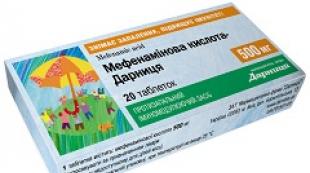
As soon as the acute phase subsides, deworming is carried out. To combat liver fluke, select the following medications marked in the table.
 Conservative treatment is supervised by a doctor. The effectiveness of the drugs given as examples reaches 90%. Albendazole and Nemozol are practically not used for deworming purposes due to their low effectiveness.
Treatment of the disease in the chronic stage
To combat running form For fascioliasis, antispasmodics and physiotherapy are widely used. After alleviating the pain syndrome and eliminating problems with the patency of the biliary tract, the specialist selects anthelmintics, in particular, preference is given to Chloxyl.
The prognosis for recovery in the initial and acute stages of the pathology is optimistic, which cannot be said about advanced invasion. In the latter case, even after complete expulsion of trematodes from the body, symptoms of liver damage continue to haunt the person.
Besides medication course treatment, in the treatment of any helminthiasis it is recommended to adhere to certain nutritional principles.

Traditional treatment
I would like to note right away that fascioliasis is an insidious disease. That's why Alternative medicine has little chance of dealing with it on his own. Despite this, recipes to combat fascioliasis exist. It is advisable to discuss their use with your attending physician, who may decide to include them in the complex of his work.
The first recipe. Fresh leaves 1 kg of sorrel pour into a liter silicon water, place in a boiling bath and heat for about 2 hours. Strain the finished broth, squeeze the raw materials into it, sweeten 50 g granulated sugar and drink one sip throughout the day, regardless of meals. This one is prohibited folk method pregnant and nursing mothers, persons with urinary and cholelithiasis.
The second recipe.
Brew wolfberry flowers with boiling water in a ratio of 1:50. Take the cooled infusion ½ teaspoon 3 times a day before meals. It must be remembered that the plant is poisonous, so it is important to follow the recommendations for preparing medicinal preparations from it.
Recipe three.
Art. Brew a spoonful of centaury with 300 ml of boiling water, cover and leave for half an hour. Take the finished infusion by sip 30 minutes before meals.
- Complications
- Fascioliasis can cause consequences such as:
- purulent cholangitis;

- cirrhosis;
- liver abscess;
mechanical jaundice;
damage to the mammary glands, lungs, brain. In all these cases, specific surgical treatment is required. This disease, referred to by experts as chronic zoonotic biohelminthiasis, causes enormous damage every year
agriculture . In addition to the death of a large number of livestock, milk yields from cows are reduced, and the quality of wool from goats and sheep deteriorates. The liver of infected livestock becomes unfit for food. The disease is common in Russia, Kazakhstan and Central Asia. Cattle become infected with fascioliasis on pastures where there is access to standing water and by eating grass clippings. Trematode larvae localized in these places penetrate the animal’s body and provoke disease.

Incubation period
lasts about 30 days. And then the acute phase of fascioliasis in animals appears. During it, a high temperature rises, appetite disappears and vomiting develops. As soon as these symptoms appear, the animal must be thoroughly examined. If this is not done, helminthiasis will become chronic. The veterinarian, after confirming fascioliasis in ruminants, prescribes anthelmintic and
symptomatic therapy
in the form of hepatoprotectors and antihistamines.
Not only cattle, but also pigs suffer from the disease. They have the same symptoms and complications that are characteristic of fascioliasis in cows and sheep. That is, treatment of pork trematode is carried out in the same way.
- Prevention Prevention of infection by invasion is based on the following measures: no hit exception boiled water from
- natural sources

- into the body; .
Taxonomy. Fascioliasis in humans is caused by two types of helminths: Fasciola hepatica (from the Latin fasciola - liver bandage) and Fasciola gigantica (giant fluke) of the order Fasciolidida, family Fasciolidae.
Morphology and life cycle of development. The common liver fluke has a flat leaf-shaped body 30-50 mm long and 8-12 mm wide. Its front part is covered with spines and extended into a proboscis, on which the oral and ventral suckers are located. The oral sucker contains the oral opening leading to the pharynx, followed by the esophagus. Two branches of the intestine with many branching lateral processes extend from the esophagus.
In the anterior part of the body, following the abdominal sucker, there is a compactly located rosette-shaped uterus, the loops of which are filled with eggs. Next are the branched ovaries and testes.
The eggs of F. hepatica, measuring 0.13-0.14x0.07-0.09 mm, are yellowish-brown in color with an operculum and a thickened shell at the poles (Fig. 27). Fasciola gigantica has an elongated shape (33-76x5-12 mm). The eggs of the giant fasciola are brown in color, measuring 0.15-0.19x0.07-0.09 mm.
The definitive hosts of fasciolae are humans, small and large livestock, horses, and pigs; intermediate -
freshwater shellfish. Fasciola eggs with feces fall into environment, where, depending on temperature and humidity, their development occurs in periods from 4-6 weeks to several months. At the first stage life cycle they develop into cilia-covered larvae called miracidia.
When they get into the water, they quickly penetrate into mollusks, where a complex asexual reproduction with the sequential formation of sporocysts, giving rise to generations of redia larvae (on behalf of F. Redi, who refuted the theory of spontaneous generation), and the latter into caudate cercariae larvae. Floating in water, cercariae attach to the stems of underwater plants and turn into invasive larvae - adolescaria, which are cercariae covered with a shell (from the Latin adolesco - grow + + (cer) carium).
On plants and in moist soil they remain viable for up to two years. The larvae enter the intestine of the final host with water or plants, then penetrate into the wall small intestine and carried by the bloodstream to the liver. The process of maturation, laying and release of eggs in the body of the definitive host takes more than 2 months, and the entire development cycle of the helminth takes at least 4-5 months.
Clinic and epidemiology. Fascioliasis is a zoonotic disease affecting the biliary system. The source of invasion for humans is agricultural and
|
||||||||||||||||||||||||||||||||||||||||||||||||||||||||||||||||||||||||||||||||||||||||||||||||||||||||
|
||||||||||||||||||||||||||||||||||||||||||||||||||||||||||||||||||||||||||||||||||||
 |
||||||||||||||||||||||||||||||||||||||||||||||||||||||||||||||||||||||||||||||||||||
wild animals.
The incubation period for fascioliasis is 1-8 weeks.
There are acute and chronic phases of its development. The acute form of the disease begins without prodromal period with a sudden rise in temperature to 38-39 ° C, pain in the right hypochondrium, often of a paroxysmal nature. The liver is significantly enlarged and painful. During the first day, jaundice or subicteric sclera may appear, and polymorphic rashes may appear on the skin. Typically, fever with fascioliasis lasts from 2-5 days to 2-3 weeks. Then fascioliasis enters a chronic phase, in which there are two main clinical variant: chronic uncomplicated fascioliasis and chronic fascioliasis complicated by bacterial infection of the biliary tract.
Uncomplicated fascioliasis occurs in waves, with periodic exacerbations and remissions, is characterized mainly by dyspeptic disorders (decreased appetite, nausea, vomiting), moderate pain syndrome. There is no fever or significant changes in the blood picture. In some patients, during the period of exacerbation, pain in the upper abdomen of a girdle nature is noted. During the period of remission, the condition of the patients is not impaired, their working capacity is preserved.
Chronic fascioliasis, complicated by a bacterial infection of the biliary tract (myxtinvasis), occurs without remission, pain in the right hypochondrium is paroxysmal in nature and is accompanied by fever. Fascioliasis, which is caused by F. hepatica, is widespread among many species of domestic and wild animals. The most affected animals are sheep, goats, and cattle, and less commonly, pigs, horses, and dogs. In humans, this invasion usually occurs sporadically in temperate latitudes, but in the tropical zone of Asia, Africa, Latin America The incidence of fascioliasis can reach a high level. Human infection with F. gigantica has been described in Vietnam, the Hawaiian Islands and several African countries.
The main factors for the transmission of fascioliasis are water, edible greens growing in reservoirs, on wet or irrigated lands; vegetables and fruits washed with water contaminated with helminth larvae. The risk of human infection increases in hot conditions humid climate in areas with an abundance of small bodies of water - mollusk biotopes.
Diagnosis. Diagnosis is difficult due to meager quantity eggs excreted in feces. As a result of this, studies are repeated many times; more often, fasciolae eggs are found in the duodenal contents. They can occasionally be found in the feces of patients who have eaten livestock liver infested with fascioli, but these so-called “transit eggs” of the helminth pass through intestinal tract without causing illness.
In the acute phase of fascioliasis, the diagnosis is confirmed using serological reactions, in particular enzyme immunoassay.
Forecast. When treating fascioliasis in the acute phase of the disease, the prognosis is quite favorable.
Prevention and treatment. Prevention is based on streamlining the water supply system and improving the sanitary and hygienic culture of the population. When drinking water from stagnant or weakly flowing reservoirs for drinking and household needs, it must be boiled. In the fight against fascioliasis in farm animals, it is recommended to deworm them in the spring to protect pastures from contamination by fasciola eggs.
For the treatment of fascioliasis, triclabendazole, bithionol and chloroxyl are prescribed. When found bacterial microflora V biliary tract antibiotics are used with a preliminary determination of the pathogen's sensitivity to them.
The causative agent of fascioliasis is the liver fluke (fasciolas hepatica sand gigantic) flatworm from the genus trematodes. There are 2 types of different sizes. The giant fluke reaches 7 cm, and the liver fluke - 2–3 cm. The main host of the liver fluke is domestic animals - sheep, cows and goats.
Fasciola hepatica eggs live at the bottom of ponds, lakes and rivers
Fasciola eggs get into the animal's feces fresh water, where after 1–2 weeks the larvae emerge. They are swallowed by snails, within which the larvae mature for several weeks. Mature larvae return to the reservoir, but are already covered with a capsule, with the help of which they attach to plants or float in the water.
People and animals become infected with fascioliasis by ingesting water from a pond.
In the intestines of humans and animals, the larva emerges and first pierces the intestinal wall, then the peritoneum and then the liver, while trying to get into bile ducts. Here they ripen and remain for many years. The full cycle of fasciola migration in humans lasts 3–4 months. Having settled in the bile ducts, fasciola lays eggs, which are excreted in the feces.
Infection with liver and giant flukes occurs when drinking water from a pond or plants on the shore, as well as fruits and vegetables washed with this water. Infection of people with fascioliasis also occurs when they consume insufficiently heat-treated liver of infected animals.
Fascioliasis occurs in acute and chronic form. Pathogenesis acute form The disease is initially caused by mechanical damage to the liver tissue during the introduction of fasciola into its parenchyma. Once in the liver and bile ducts, fascioli secrete toxic substances that cause an allergic reaction in humans.

The mechanism of infection is alimentary, and the route is food or water
Signs of fascioliasis
From the moment of infection to the first symptoms of fascioliasis, it takes from 1 week to several months. This is due to the number of larvae ingested and the reaction immune system person. In half of cases, fascioliasis in humans is asymptomatic. In other patients, symptoms of the disease appear when the fascioli pierce the liver during migration. During this period, the following symptoms appear:
- pain in the right hypochondrium or non-localized;
- temperature increase to 39.0–40.0 °C;
- yellowness of the skin;
- dry cough;
- itchy, red, swollen rash;
- paroxysmal headaches;
- Nausea and vomiting are a variable symptom.
Acute symptoms of human fascioliasis last from 2 to 6 months and stop after fasciola enters the bile ducts.
In the chronic stage of fascioliasis allergic reaction also appears in the form of a rash, but predominates gastrointestinal signs diseases. Chronic course Fascioliasis in humans is manifested by symptoms of cholecystitis, cholangitis or pancreatitis. In this case, there are paroxysmal pains in the right hypochondrium, accompanied by fever, nausea and vomiting. Yellowness of the skin and sclera appears periodically. Accession bacterial infection accompanied by hepatic colic. When examining the patient, an enlargement of the liver and spleen is revealed.
The diagnosis of fascioliasis is established on the basis of an epidemiological history, symptoms of the disease and laboratory examination. Diagnostic methods:

When liver fluke eggs are detected in feces, the diagnosis of fascioliasis is beyond doubt
IN acute period disease applies complex treatment which involves preparing the patient for use anthelmintic drugs. At the beginning of the course, choleretic and sorbents are used.
Treatment regimen:
- Antihistamines.
- Intravenous administration of calcium chloride.
- For the specific treatment of fascioliasis, Triclabendazole is used once at a dose of 10 mg per 1 kg of human weight.
- An alternative drug, Bitinolol, is used for treatment.
- Chloxil is taken 3 times a day at the rate of daily dose 60 mg per 1 kg of patient weight.
- In the absence of these drugs, Praziquantel is also used to treat fascioliasis. However, its effectiveness is somewhat inferior.
- Biltricide is used at a dose of 60 mg per 1 kg of body weight. The drug is taken in 2 doses.
- The addition of a bacterial infection requires the use of antibiotics.

Drugs for fascioliasis are prescribed from the group of anthelmintics
After a course of treatment, blood monitoring for antibodies is necessary, because the effectiveness of the drugs is 80%. If the antibody titer does not decrease, a repeat course treatment. After 6 months, feces and duodenal contents are examined for worm eggs.
How to prevent the disease
Effective measures to prevent disease are the development of hygiene habits:
- washing hands after every stay outside the home;
- a ban on the use of water from a reservoir for washing hands, fruits or vegetables;
- never drink water from a reservoir;
- do not use plants from the shore of the reservoir for food;
- Do not consume thermally insufficiently processed animal liver.
Control of large and small livestock is also a measure to prevent infection.


