Human structure. Connections of the bones of the upper limb
At the free part upper limb highlight the joints of the scapula, humerus, bones of the forearm and hand (Table 13).
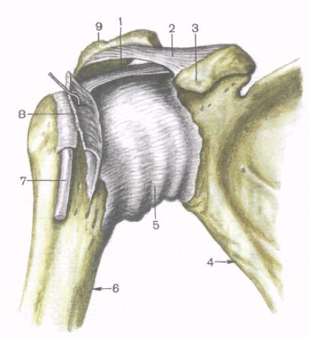
Shoulder joint (art. humeri) is formed by the glenoid cavity of the scapula and the head of the humerus (Fig. 100, 101). The articular surface of the head is spherical, almost 3 times larger than the flat surface of the glenoid cavity of the scapula. The articular cavity is supplemented along the edges of the cartilaginous labrum(labrum glenoidale), which increases the congruence of the articular surfaces and the capacity of the articular fossa. The joint capsule is attached to outside articular labrum, as well as anatomical neck humerus. The capsule of the shoulder joint is thin, weakly stretched, and loose. On top, the articular capsule is strengthened by the only one in this joint coracobrachial ligament(lig. coracohumerаle), which begins at the base coracoid process scapula and is attached to the upper part of the anatomical neck of the humerus. The fibers of the tendons of adjacent muscles (subscapularis, etc.) are also woven into the capsule. The synovial membrane of the joint capsule forms two protrusions. One of them - intertubercular synovial vagina(vagina synovialis intertubercularis) like a sheath surrounds the tendon of the long head of the biceps brachii muscle, passing through the articular cavity. Second protrusion - subtendinous bursa of the subscapularis muscle(bursa subtendinea m. subscapularis) is located at the base of the coracoid process, under the tendon of this muscle.
The shape of the articular surfaces of the shoulder joint is spherical. It has a large range of motion around three axes, which is facilitated by a loose joint capsule, a big difference in the size of the articulating surfaces, the absence of powerful ligaments. Flexion and extension occur around the frontal axis. The total range of these movements is approximately 120°. Relatively sagittal axis abduction is performed (up to horizontal level) and casting hands. Range of movements up to 100". In relation to vertical axis rotations outward (supination) and inward (pronation) are possible with a total volume of up to 135°. The shoulder joint also undergoes circular movements (circumduxio). Movement of the upper limb above the horizontal level is performed in the sternoclavicular joint when raising the scapula together with the free upper limb.
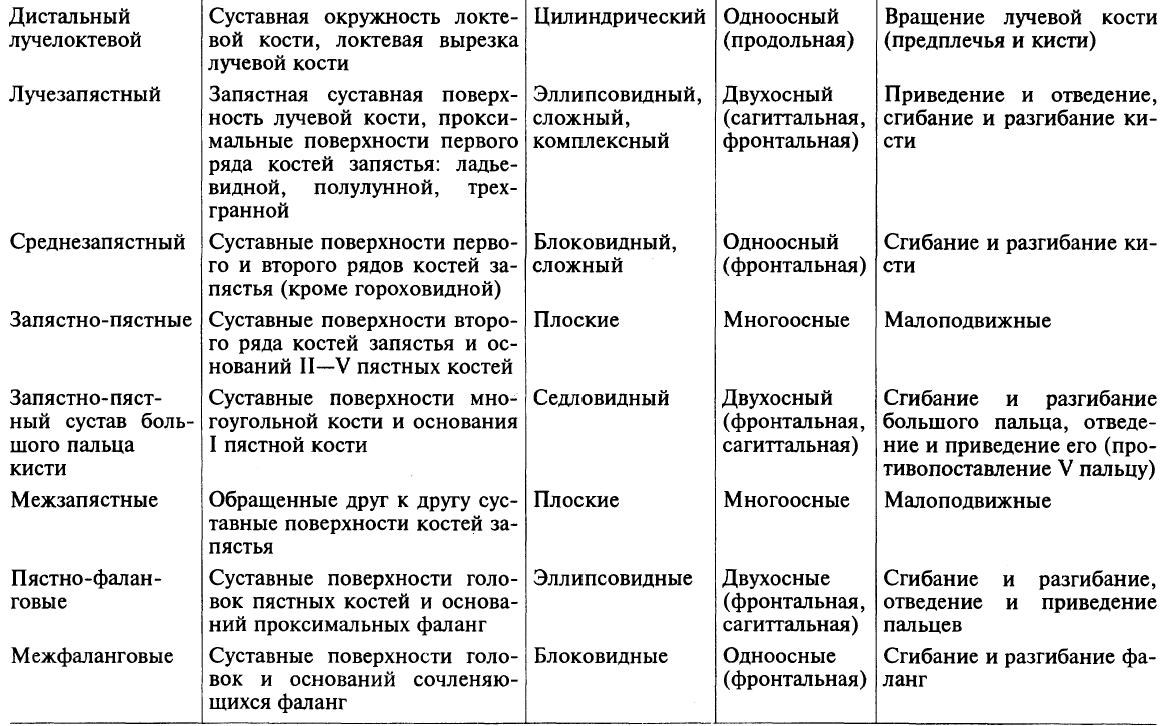
Rice. 100. Shoulder joint; front view. 1 - coracohumeral ligament; 2 - coracoacromial ligament; 3 - coracoid process; 4 - blade; 5 - articular capsule; 6 - humerus; 7 - biceps brachii tendon ( long head); 8 - tendon of the subscapularis muscle; 9 - acromion.
Rice. 101. Shoulder joint. (Cut in the frontal plane.) 1 - coracoid process; 2.5 - tendon of the biceps brachii muscle (long head); 3 - articular cavity; 4 - articular capsule; 6 - intertubercular synovial vagina; 7 - head of the humerus; 8 - coracohumeral ligament.
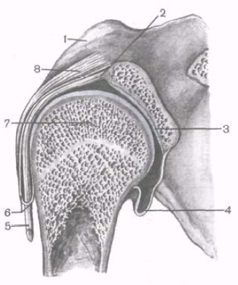
An x-ray of the shoulder joint (Fig. 102) clearly identifies the head of the humerus and the glenoid cavity of the scapula. The contours of the inferomedial part of the head overlap the glenoid cavity of the scapula. The X-ray slit in the image looks like an arcuate strip.
Elbow joint(art. cubiti) is formed by three bones: humerus, radius and ulna (Fig. 103, 104). The bones form three joints, enclosed in a common joint capsule.
Shoulder-ulnar joint
(art. humeroulnaris) trochlear, formed by the connection of the trochlea of the humerus and the trochlear notch ulna.
Rice. 102. X-ray of the shoulder joint, left. 1 - spine of the scapula; 2 - acromion; 3 - coracoid process; 4 - collarbone; 5 - head of the humerus; 6 - greater tubercle (humerus); 7 - first rib; 8 - x-ray joint space; 9 - blade; 10 - humerus.
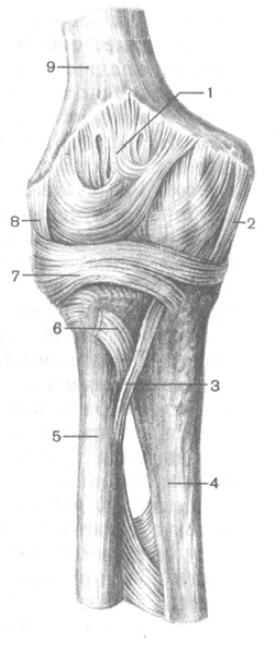
Rice. 103. Elbow joint; front view. 1 - articular capsule; 2 - ulnar collateral ligament; 3 - oblique chord; 4 - ulna; 5 - radius; 6 - biceps brachii tendon (cut off); 7 - ring ligament radius; 8 - radial collateral ligament; 9 - humerus.
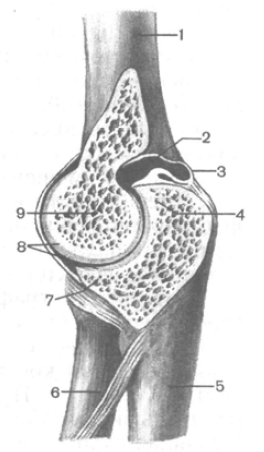
Rice. 104. Elbow joint. (Cut in the sagittal plane.) 1 - humerus; 2 - articular cavity; 3 - articular capsule; 4 - olecranon; 5 - ulna; 6 - radius; 7 - coronoid process; 8 - articular cartilage; 9 - block of the humerus.
Humeral joint(art. humeroradialis) spherical, represents the articulation of the head of the humerus and the articular cavity of the radius.
Proximal beam elbow joint (art. radioulnaris proximalis) - cylindrical in shape, formed by the articular circumference of the radius and the radial notch of the ulna. The general joint capsule is free. On the humerus, the articular capsule is attached relatively high above the articular cartilage of the humerus block, so the coronoid and radial fossae and the olecranon fossa are located in the joint cavity. The lateral and medial epicondyles of the humerus are located outside the articular cavity. On the ulna, the articular capsule is attached below the edge of the articular cartilage of the coronoid process and at the edge of the trochlear notch of the olecranon process. On the radius, the capsule is attached to its neck. The joint capsule is strengthened by ligaments. Ulnar collateral ligament(lig. collaterale ulnare) originates below the edge of the medial epicondyle of the humerus, expands in a fan-shaped manner and attaches along the entire medial edge of the trochlear notch of the ulna. Radial collateral ligament(lig. collaterale radiale), starting at the lower edge of the lateral epicondyle of the humerus, is divided into two bundles. The anterior bundle covers the neck of the radius from the front and is attached at the anterior outer edge of the trochlear notch of the ulna. Posterior bun This ligament covers the neck of the radius from the back and is woven into the annular ligament of the radius. Annular ligament of the radius(lig. annulare radii) begins at the anterior edge of the radial notch of the ulna, covers the neck of the radial bone in the form of a loop and is attached to the posterior edge of the radial notch. Between the distal edge of the radial notch of the ulna and the neck of the radius is located quadrate ligament(lig. quadratum).
In the elbow joint, movements around the frontal axis are possible - flexion and extension of the forearm with a total volume of up to 170°. When flexing, the forearm deviates slightly medially and the hand rests not on the shoulder, but on the chest. This is due to the presence of a notch on the trochlea of the humerus, which promotes helical displacement of the forearm and hand. Around the longitudinal axis of the radius in the proximal radioulnar joint, the radius rotates together with the hand. This movement occurs simultaneously in both the proximal and distal radioulnar joints.
When X-raying the elbow joint in a lateral projection (the forearm is flexed 90°), the line of the X-ray joint space is limited by the trochlear notch of the ulna and the head of the radius on one side and the condyle of the humerus on the other. In direct projection, the x-ray joint space is zigzag-shaped and has a thickness of 2-3 mm. The joint space of the proximal radioulnar joint is also visible.
Rice. 105. Connection of the bones of the forearm, right; front view. 1 - ulna; 2 - styloid process of the ulna; 3 - articular disc; 4 - styloid process of the radius; 5 - interosseous membrane of the forearm; 6 - radius; 7 - tendon of the biceps brachii; 8 - annular ligament of the radius.
The bones of the forearm are connected using discontinuous and continuous connections (Fig. 105). Continuous connection is interosseous membrane of the forearm(membrana interossea antebrachii).
It is a strong connective tissue membrane stretched between the interosseous edges of the radius and ulna. Down from the proximal radioulnar joint, a fibrous cord is visible between both bones of the forearm - oblique chord(chorda obliqua).
Discontinuous joints include the proximal radioulnar joint (discussed above) and the distal radioulnar joint, as well as the hand joints.
Distal radioulnar joint(art. radioulnaris distalis) is formed by the connection of the articular circumference of the ulna and the ulnar notch of the radius. This joint is separated from wrist joint articular disc(discus articularis), located between the ulnar notch of the radius and the styloid process of the ulna. The articular capsule of the distal radioulnar joint is free, attached to the edge of the articular surfaces and the articular disc. The capsule usually protrudes proximally between the bones of the forearm, forming pouch-shaped recess(recessus sacciformis).
The proximal and distal radioulnar joints functionally together form a combined cylindrical joint with a longitudinal axis of rotation (along the forearm). In these joints, the radius bone, together with the hand, rotates around the ulna. In this case, the proximal epiphysis of the radius rotates in place as the head of the radius is held in place by the annular ligament of the radius. The distal epiphysis of the radius describes an arc around the head of the radius, which remains motionless. The average range of rotation in the radioulnar joints (supination and pronation) is approximately 140°.
The joints of the free upper limb connect the bones of this part to each other, as well as to the girdle of the upper limb. Shoulder joint(articulatio humeri) is formed by the head of the humerus, the articular cavity of the scapula, which is complemented by the articular lip. The joint capsule covers the head of the humerus on the anatomical neck, and on the scapula it is attached along the edge of the glenoid cavity. The joint is strengthened by the coracobrachial ligament and muscles. The tendon of the long head of the biceps brachii muscle passes through the joint cavity. The shoulder joint is a ball-and-socket joint in which movement is possible around three axes: frontal, sagittal and vertical. Elbow joint(articulatio cubiti) - complex, it includes the humeroulnar, humeroradial and proximal radioulnar joints. These three joints share a common joint capsule, which is strengthened by the radial and ulnar collateral ligaments, as well as the annular ligament of the radius. The elbow joint is a trochlear joint: it allows flexion, extension and rotation of the forearm. Distal radioulnar joint(articulatio radioulnaris distalis) is an independent joint, and the proximal radioulnar joint is included in the elbow joint. However, they form a single combined cylindrical (rotational) joint. If the rotation of the radius occurs around the longitudinal axis together with the palmar surface of the hand inward, then such a movement is called pronation, and vice versa - supination. Wrist joint(articulatio radiocarpalis) is a complex ellipsoidal joint formed by the carpal articular surface of the radius and three bones of the first row of the wrist. Two types of movement are possible in it: adduction and abduction, flexion and extension, as well as a small passive circular movement. The joint is surrounded by a common capsule and is strengthened by powerful ulnar, radial, palmar and dorsal wrist ligaments. Joints of the hand include the intermetacarpal, carpometacarpal, metacarpophalangeal and interphalangeal joints. These joints are strengthened by short interosseous ligaments, which are located on the palmar and dorsal surfaces of the hand outside the joint cavities. The carpometacarpal joint has a special structure thumb. It is saddle-shaped in shape and is characterized by two types of movement: flexion and extension, adduction and abduction, possibly a circular movement, as well as opposition of the thumb to the rest. The metacarpophalangeal joints are spherical, and the interphalangeal joints are block-shaped. The structural features of the bones and joints of the hand determine its extreme mobility, which allows you to perform very subtle and varied movements.
16. Bones of the pelvic girdle and their connection.
Belt lower limb(cingulum membri inferioris) consists of a paired pelvic bone. The pelvic bone, os coxae, refers to flat bones and performs the function of movement (participation in articulations with the sacrum and thigh), protection (pelvic organs) and support (transferring the weight of the entire overlying part of the body to the lower limbs). The latter function predominates, which determines the complex structure of the pelvic bone and its fusion of three separate bones - the ilium, os ilium, pubis, os pubis, and ischium, os ischii. Fusion of these bones occurs in the area heaviest load, namely in the area of the acetabulum, which is the articular fossa hip joint, in which the articulation of the lower limb belt with the free lower limb occurs.
The ilium lies upward from the acetabulum, the pubis lies downward and anteriorly, and the ischium lies downward and posteriorly. In persons under 16 years of age, the listed bones are separated from each other by cartilaginous layers, which ossify in an adult, i.e. synchondrosis turns into synostosis.
Thanks to this, three bones become one, which has great strength, necessary to support the entire body and head. The acetabulum, acetabulum (vinegar, from acetum - vinegar), is placed on the outer side of the pelvic bone and serves to articulate with the head of the femur. Having the shape of a rather deep rounded fossa, it is delimited along the circumference by a high edge, which on its medial side is interrupted by a notch, incisura acetabuli. Articular smooth surface acetabulum has the shape of a crescent, facies lunata, while the center of the cavity, the so-called fossa acetabuli, and the part closest to the notch are rough. Ilium
The ilium, os ilium, with its lower short thick section, called the body, corpus ossis ilii, merges with the rest of the pelvic bone in the region of the acetabulum; its upper, expanded and more or less thin part forms the wing of the ilium, ala ossis ilii. The relief of the bone is determined mainly by muscles, under the action of which ridges, lines and spines were formed in places of tendon attachment, and pits in places of fleshy attachment. Thus, the upper free edge of the wing represents a thickened, S-shaped crest, crista iliaca, to which three broad abdominal muscles are attached. The ridge in front ends with the anterior superior spine, spina iliaca anterior superior, and at the rear with the posterior superior spine, spina iliaca posterior superior. Below each of these spines, on the anterior and posterior edges of the wing there is another spine: spina iliaca anterior inferior and spina iliaca posterior inferior. The lower awns are separated from the upper ones by notches. Below and anterior to the anterior inferior spine, at the junction of the ilium and the pubis, there is the iliopubic eminence, eminentia iliopubica, and downward from the posterior inferior spine lies the deep greater sciatic notch, incisura ischiadica major, which closes further downward with the ischial spine, spina ischiadica, already located on the ischium. The inner surface of the wing of the ilium is smooth, slightly concave and forms the iliac fossa, fossa iliaca, which arose in connection with the maintenance of the insides when the body is in an upright position. Posterior and inferior to the latter lies the so-called ear-shaped articular surface, facies auricularis, the place of articulation with the sonominal surface of the sacrum, and posterior and superior to the articular surface there is a tuberosity, tuberositas iliaca, to which the interosseous sacroiliac ligaments are attached. The iliac fossa is separated from the inner surface of the underlying body of the ilium by an arched edge called linea arcuata. On the outer surface of the wing of the ilium, rough lines are visible, sometimes more or less clearly - traces of the attachments of the gluteal muscles (lineae gluteae anterior, posterior et inferior). pubic bone
The pubic bone, os pubis, has a short thickened body, corpus ossis pubis, adjacent to the acetabulum, then upper and lower branches, ramus superior and ramus inferior ossis pubis, located at an angle to each other. At the vertex of the angle facing the midline there is oval shape surface, facies symphysialis, junction with the pubic bone of the other side. 2 cm lateral from this surface there is a small pubic tubercle, tuberculum pubicum, from which the pubic crest, pecten ossis pubis, extends along the posterior edge of the upper surface of the ramus superior, passing further posteriorly into the above-described linea arcuata of the ilium. On the lower surface of the superior branch of the pubic bone there is a groove, sulcus obturatorius, the site of passage of the obturator vessels and nerve. Ischium
The ischium, os ischii, like the pubis, has a body, corpus ossis ischii, which is part of the acetabulum, and a branch, ramus ossis ischii, forming an angle with each other, the apex of which is greatly thickened and represents the so-called ischial tubercle, tuber ischiadicum. Along the posterior edge of the body, upward from ischial tuberosity, the lesser sciatic notch, incisura ischiadica minor, is located, separated by the ischium, spina ischiadica, from the greater sciatic notch, incisura ischiadica major. The branch of the ischium, moving away from the ischial tuberosity, then merges with the lower branch of the pubis. As a result, the pubic and ischium their branches surround the obturator foramen, foramen obturatum, which lies inferiorly and medially from the acetabulum and has the shape of a triangle with rounded corners.
As a result, all types of connections are observed in the human pelvis, reflecting successive stages skeletal development: synarthrosis in the form of syndesmoses (ligaments), synchondrosis (between individual parts of the pelvic bone) and synostosis (after their fusion into pelvic bone), symphysis (pubic) and diarthrosis (sacroiliac joint). The overall mobility between the pelvic bones is very small (4 - 10 degrees).
1. Sacroiliac joint, art. sacroiliaca, belongs to the type of tight joints (amphiarthrosis), formed by the ear-shaped articular surfaces of the sacrum and ilium in contact with each other. It is strengthened by ligg. sacroiliaca interossea, located in the form of short bundles between the tuberositas iliaca and the sacrum, which are one of the strongest ligaments of all human body. They serve as the axis around which movements of the sacroiliac joint occur. The latter is also strengthened by other ligaments connecting the sacrum and ilium: front - ligg. sacroiliaca ventralia, behind - ligg. sacroiliaca dorsalia, as well as lig. iliolumbale, which extends from transverse process V lumbar vertebra to crista iliaca.
The sacroiliac joint is vascularized from aa. lumbalis, iliolumbalis et sacrales laterales. The outflow of venous blood occurs into the veins of the same name. The outflow of lymph is carried out through deep lymphatic vessels in nodi lymphatici sacrales et lumbales. The innervation of the joint is provided by the branches of the lumbar and sacral plexuses.
2. The pubic symphysis, symphysis pubica, connects, located in the midline, both pubic bones to each other. Between the facies symphysialis of these bones facing each other, covered with a layer of hyaline cartilage, there is a fibrocartilaginous plate, discus interpubicus, in which usually, starting from the age of 7, there is a narrow synovial cleft (half-joint). The pubic symphysis is supported by dense periosteum and ligaments; on the upper edge - lig. pubicum superius and on the lower - lig. arcuatum pubis; the latter smoothes the angle under the symphysis, angulus subpubicus.
3. Lig. sacrotuberale and lig. sacrospinale - two strong interosseous ligaments connecting the sacrum with the pelvic bone on each side: the first - with tuber ischii, the second - with spina ischiadica.
The described ligaments complement the bony skeleton of the pelvis in its posteroinferior section and transform the greater and lesser sciatic notches into the openings of the same name: foramen ischiadicum majus et minus.
4. The obturator membrane, membrana obturatoria, is a fibrous plate that covers the foramen obturatum of the pelvis, with the exception of the superolateral corner of this opening.
Attaching to the edges of the sulcus obturatorius of the pubic bone located here, it turns this groove into the canal of the same name, canalis obturatorius, caused by the passage of the obturator vessels and nerve.
The skeleton of the upper limbs is divided into two sections: the skeleton of the upper limb girdle ( shoulder girdle) and the skeleton of the free upper limb (Fig. 36).
Bones of the upper limb girdle
The skeleton of the upper limb girdle is formed by two paired bones: the scapula and the clavicle.
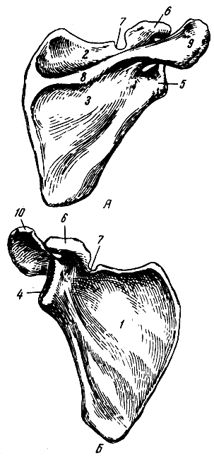
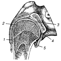
The scapula (scapula) is a flat bone (Fig. 37), on which there are two surfaces (costal and dorsal), three edges (upper, medial and lateral) and three angles (lateral, upper and lower). The lateral angle is thickened and has a glenoid cavity for articulation with the humerus. Above the glenoid cavity is the coracoid process. The costal surface of the scapula is slightly concave and is called the subscapular fossa; the muscle of the same name begins from it. The dorsal surface of the scapula is divided by the spine of the scapula into two fossae - supraspinatus and infraspinatus, in which the muscles of the same name lie. The spine of the scapula ends with a protrusion - the acromion (humeral process). It has an articular surface for articulation with the collarbone.
Collarbone(clavicula) - an S-shaped curved bone with a body and two ends - sternum and acromial (see Fig. 35). The sternal end is thickened and connects to the manubrium of the sternum. The acromial end is flattened and connects to the acromion of the scapula. The lateral part of the clavicle is convexly facing back, and the medial part is facing forward.
Bones of the free upper limb
The skeleton of the free upper limb (arm) includes the humerus, forearm bones and hand bones (see Fig. 36).
Brachial bone(humerus) - a long tubular bone, consists of a body (diaphysis) and two ends (epiphyses) (Fig. 38). At the proximal end there is a head, separated from the rest of the bone by an anatomical neck. Below the anatomical neck, on the outer side, there are two elevations: the greater and lesser tubercles, separated by the intertubercular groove. Distal to the tubercles is a slightly narrowed section of bone - surgical neck. This name is due to the fact that bone fractures occur more often in this place.
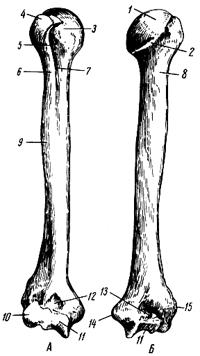
The upper part of the body of the humerus is cylindrical, and the lower part is triangular. In the middle third of the body of the humerus, a groove runs spirally at the back radial nerve. The distal end of the bone is thickened and is called the condyle of the humerus. On the sides it has protrusions - the medial and lateral epicondyles, and below are the head of the condyle of the humerus for connection with the radius and the block of the humerus for articulation with the ulna. Above the block in front there is a coronoid fossa, and behind there is a deeper fossa of the olecranon process (the processes of the same name of the ulna enter into them).
Bones of the forearm: the radial one is located laterally, the ulnar one occupies a medial position (Fig. 39). They are long tubular bones.
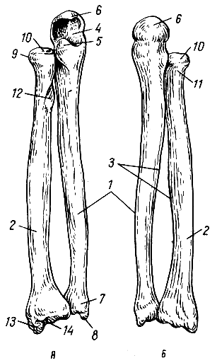
Radius(radius) consists of a body and two ends. At the proximal end there is a head, and on it there is an articular fossa, with the help of which the radius articulates with the head of the condyle of the humerus. The head of the radius also has an articular circle for connection with the ulna. Below the head is the neck, and below it is the tuberosity of the radius. There are three surfaces and three edges on the body. The sharp edge faces the edge of the ulna of the same shape and is called the interosseous. At the distal extended end of the radius there is a carpal articular surface (for articulation with the proximal row of carpal bones) and an ulnar notch (for articulation with the ulna). Outside at the distal end there is a styloid process.
Elbow bone(ulna) consists of a body and two ends. At the thickened proximal end there are coronoid and olecranon processes; they are limited by the trochlear notch. On the lateral side at the base of the coronoid process there is the radial notch. Below the coronoid process there is a tuberosity of the ulna.
The body of the bone is triangular in shape, and there are three surfaces and three edges on it. The distal end forms the head of the ulna. The surface of the head facing the radius is rounded; there is an articular circle on it for connection with the notch of this bone. WITH medial side the styloid process extends down from the head.
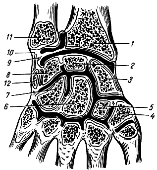
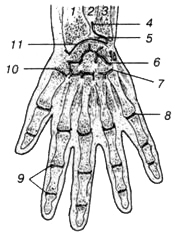
Hand bones are divided into carpal bones, metacarpal bones and phalanges (fingers) (Fig. 40).
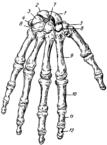
Carpal bones- ossa carpi (carpalia) are arranged in two rows. The proximal row consists of (in the direction from the radius to the ulna) the scaphoid, lunate, triquetrum, and pisiform bones. The first three are arched, forming an ellipsoidal surface for connection with the radius. The distal row is formed the following bones: trapezium, trapezoid, capitate and hamate bones.
The bones of the wrist do not lie in the same plane: on the back they form a convexity, and on the palmar side they form a concavity in the form of a groove - the groove of the wrist. This groove is deepened medially by the pisiform bone and the hook of the hamate, and laterally by the tubercle of the trapezium bone.
Metacarpal bones five in number are short tubular bones. Each of them has a base, a body and a head. The bones are counted from the side of the thumb: I, II, etc.
Phalanges of fingers belong to tubular bones. The thumb has two phalanges: proximal and distal. Each of the remaining fingers has three phalanges: proximal, middle and distal. Each phalanx has a base, a body and a head.
Connections of the bones of the upper limb
Sternoclavicular joint(articulatio sternoclavicularis) is formed by the sternal end of the clavicle with the clavicular notch of the manubrium of the sternum. Inside the joint cavity is an articular disc, which divides the joint cavity into two parts. The presence of a disc allows movement in the joint around three axes: sagittal - movement up and down, vertical - forward and backward; Rotational movements are possible around the frontal axis. This joint is strengthened by ligaments (interclavicular, etc.).
AC joint(articulatio acromiclavicularis) formed by the acromial end of the clavicle and the acromion of the scapula, flat in shape; the movements in it are insignificant.
Shoulder joint(articulatio humeri) is formed by the head of the humerus and the articular cavity of the scapula (Fig. 41), supplemented along its edge by an articular lip. The joint capsule is thin. In her top part fibers of the coracobrachial ligament are woven. The joint is strengthened mainly by muscles, especially the long head of the biceps muscle, the tendon of which passes through the joint cavity. In addition, the extra-articular coracoacromial ligament takes part in strengthening the joint - a kind of arch that prevents the abduction of the arm in the joint above the horizontal line. Abduction of the arm above this line is carried out due to movement in the shoulder girdle.
The shoulder joint is the most mobile joint in the human body. Its shape is spherical. It allows movements around three axes: frontal - flexion and extension; sagittal - abduction and adduction; vertical - rotation. In addition, circular motion is possible at this joint.
Elbow joint(articulatio cubiti) is formed by three bones: the distal end of the humerus and the proximal ends of the ulna and radius (Fig. 42). There are three joints: the humeroulnar, brachioradial and proximal radioulnar. All three joints are united by a common capsule and have a common articular cavity. The joint is strengthened on the sides by the radial and ulnar collateral ligaments. The strong annular ligament of the radius runs around the head of the radius.
The humeral-ulnar joint is block-shaped in shape; flexion and extension of the forearm are possible in it. The humeral joint is ball-and-socket.
Joints of the bones of the forearm. The radius and ulna are connected through the proximal and distal radioulnar joint and the interosseous membrane (membrane) of the forearm. The radioulnar joints are formed by notches and articular circles at the corresponding ends of the bones of the forearm, with the proximal joint being part of the elbow, and the distal one having its own capsule. Both joints make up combined joint, allowing rotation of the radius around the ulna. Inward rotation is called pronation, and outward rotation is called supination. The hand rotates together with the radius.
The interosseous membrane of the forearm is located between the bodies of two bones and is attached to their interosseous edges.
Wrist joint(articulatio radiocarpea) is formed by the distal end of the radius and the proximal row of carpal bones, excluding the pisiform bone (Fig. 43). The ulna does not participate in the formation of the joint. The joint is strengthened by the radial and ulnar collateral ligaments of the wrist and ligaments running along its palmar and dorsal sides. The joint has an elliptical shape; the following movements are possible in it: flexion and extension, abduction and adduction, as well as circular movements of the hand.
Intercarpal joint formed by the distal and proximal rows of carpal bones. The joint cavity is S-shaped. Functionally, it is connected to the wrist joint; together they form the combined joint of the hand.
Carpometacarpal joints formed by the distal row of carpal bones and the base of the metacarpal bones. The first carpometacarpal joint of the thumb should be highlighted (the articulation of the trapezium bone with the first metacarpal bone). It has a saddle shape and is highly mobile. The following movements are possible in it: flexion and extension of the thumb (together with the metacarpal bone), abduction and adduction; in addition, circular movements are possible. The remaining carpometacarpal joints are flat in shape and inactive.
Metacarpophalangeal joints formed by the heads of the metacarpal bones and the bases of the proximal phalanges. These joints are spherical in shape; they allow flexion and extension, abduction and adduction of the fingers, as well as passive rotational movements.
Interphalangeal joints Block-shaped in shape, flexion and extension of the phalanges of the fingers are possible in them.
Connections of the bones of the upper limb girdle
1. Own ligaments shoulder blades- these are two ligaments that are not related to joints. The first of them - the coracoacromial - is the strongest ligament of the scapula, has the shape of a triangular plate, starts from the anterior edge of the apex of the acromial process and is widely attached to the coracoid process. It forms the “arch of the shoulder joint”, protecting the joint from above and limiting the movement of the humerus in this direction.
The second - the superior transverse ligament of the scapula - is a short thin bundle thrown over the notch of the scapula. Together with the notch of the scapula, it forms an opening for the passage of blood vessels and nerves, and often ossifies.
2. Connections between the bones of the belt. The acromioclavicular joint (articulatio acromioclavicularis) is formed between the acromion process and the collarbone. Its articular surfaces are slightly curved, less often flat. The joint capsule is tight, strengthened by the acromioclavicular ligament. Very rarely, an intra-articular disc is found in this joint, which divides the joint cavity into two floors.
Movements in the acromioclavicular joint are possible in all directions, but their volume is insignificant. In addition to the mentioned ligament, the strong coracoclavicular ligament prevents movement. It is divided into two ligaments: the quadrangular trapezoid, which lies laterally and anteriorly; and a narrower triangular conical, which is located more medially and posteriorly.
Both ligaments meet each other at an angle that is open medially and anteriorly.
3. Connections between the bones of the belt and the skeleton of the body. Between the collarbone and the manubrium of the sternum is the sternoclavicular joint (articulatio sternoclavicularis). The articulating surfaces are incongruent and covered with fibrous cartilage; their shape is very variable, often saddle-shaped. In the joint cavity there is an intra-articular disc that levels the articular surfaces of the bones that do not correspond well to each other. The shape of the joint is saddle-shaped. The clavicle makes the most extensive movements around the sagittal axis - up and down; around the vertical axis - forward and backward. Circular motion is possible around these two axes. The articular capsule is strengthened by the anterior and posterior sternoclavicular ligaments, with the exception of the lower surface, where the capsule is thin. These ligaments limit forward and backward movement.
In addition, the sternoclavicular joint is strengthened by the interclavicular and costoclavicular ligaments.
1 - articular disc; 2 - interclavicular ligament; 3 - anterior sternoclavicular ligament; 4 - collarbone; 5 - 1st rib; 6 - costoclavicular ligament; 7 - sternum
Of the bones of the shoulder girdle, only the clavicle is connected at its medial end to the skeleton of the body, so the bones of the girdle have great mobility; the movements of the scapula are directed and regulated by the clavicle, so the mechanical significance of the latter is very great.
Connections of the free upper limb
This group includes the connections of the bones of the free upper limb with the girdle of the upper limb (scapula), as well as with each other.
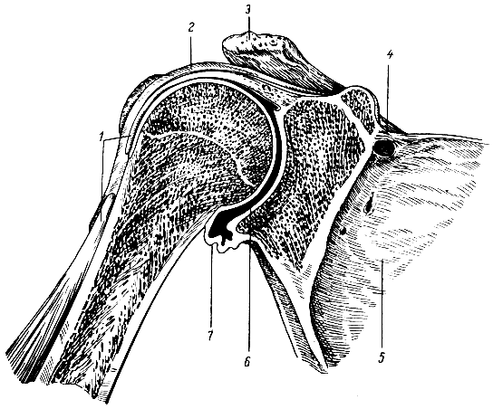
The shoulder joint (articulatio humeri) is formed by the head of the humerus and the glenoid cavity of the scapula. The articular surface of the head of the humerus is one-third (or slightly more) of the surface of the ball. The glenoid cavity is oval in shape, slightly concave and in area makes up only a quarter of the surface of the head. It is complemented by an articular lip, which increases the congruence of the articulating surfaces, which are covered with hyaline cartilage.
1 - tendon of the biceps brachii: 2 - head of the humerus; 3 - glenoid cavity of the scapula; 4 - articular lip; 5 - axillary bursa
The joint capsule is very loose; when the limb is lowered, it gathers into folds. It is attached to the shoulder blade along the edge labrum, and on the humerus - along the anatomical neck, while both tubercles remain outside the joint cavity. Spreading in the form of a bridge over the intertubercular groove, the synovial layer of the joint capsule forms a blindly ending finger-like inversion - the intertubercular synovial sheath (vagina synovialis intertubercularis) 2-5 cm long. It lies in the intertubercular groove, covering the tendon of the long head of the biceps brachii muscle, passing through the joint cavity above the head of the humerus.
The synovial membrane also forms a second permanent eversion - the subtendinous bursa of the subscapularis muscle (bursa subtendinea m. subscapularis). It is located at the base of the coracoid process of the scapula, under the tendon of the subscapularis muscle and widely communicates with the joint cavity.
In the axillary cavity, the joint capsule becomes significantly thinner and forms a permanent deep fold in which the axillary joint is located. bursa(bursa synovialis axillaris).
The capsule of the shoulder joint is thin, strengthened above and behind by the coracobrachial and articular-brachial ligaments.
- The coracobrachial ligament is well defined, starts from the base of the coracoid process and is woven into the capsule from the upper and posterior sides. The direction of its fibers almost exactly coincides with the course of the biceps tendon.
- The articular-brachial ligaments are represented by three bundles, located above and in front, intertwined with inner layer fibrous membrane of the joint capsule. They are fixed on the humerus to the anatomical neck and reach the articular labrum.
The joint capsule, in addition to the ligaments, is strengthened by the fibers of the tendons of the supraspinatus, infraspinatus, teres minor and subscapularis muscles. Consequently, the inferomedial part of the shoulder joint capsule is least strengthened.
The shape of the shoulder joint is typical spherical, multi-axial, the most mobile of all the discontinuous joints of the bones of the human body, since the articulating surfaces differ greatly in area, and the capsule is very spacious and elastic. Movements in the shoulder joint can occur in all directions. Depending on the nature of the movements, the capsule relaxes, forms folds on one side and tenses on the opposite side.
The following movements occur in the shoulder joint:
- around the frontal axis - flexion and extension;
- around the sagittal axis - abduction to the horizontal level (further movement is prevented by the arch of the shoulder, formed by two processes of the scapula with the acromiocoracoid ligament thrown between them) and adduction;
- around the vertical axis - rotation of the shoulder in and out;
- when moving from one axis to another - circular motion.
Movements around the frontal and sagittal axes are within 90°, rotation is somewhat less. Flexion, extension, and abduction of the arm almost to the vertical, performed to the maximum extent, are carried out thanks to the mobility of the scapula and additional movements in the sternoclavicular joint.
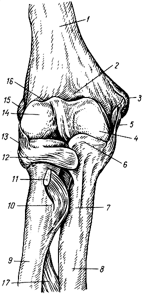
Three bones take part in the formation of the elbow joint (articulatio cubiti) - the humerus, the ulna and the radius. Three simple joints are formed between them. All three joints have a common capsule and one articular cavity, therefore, from an anatomical and surgical point of view, they are combined into one (complex) joint. All articular surfaces are covered with hyaline cartilage.
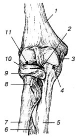
1 - humerus; 2 - proximal radioulnar joint; 3 - ulnar collateral ligament; 4 - humeral-elbow joint; 5 - ulna; 6 - interosseous membrane of the forearm; 7 - radius; 8 - tendon of the biceps brachii; 9 - annular ligament of the radius; 10 - radial collateral ligament; 11 - humeroradial joint
- Shoulder-elbow joint (articulatio humeroulnaris) formed by the articulation of the trochlea of the humerus and the trochlear notch of the ulna. The block of the humerus is a cylinder with a recess that has a screw stroke. The joint is helical or cochlear in shape, uniaxial.
- Shoulder joint (articulatio humeroradialis) is the articulation of the head of the condyle of the humerus with the articular fossa of the head of the radius. The joint is spherical in shape.
- Proximal radioulnar joint (articulatio radioulnaris proximalis) is a cylindrical joint and is formed by the articulation upper ends radius and ulna bones.
All three joints are covered by one common articular capsule. On the humerus, the capsule is attached far from the edge of the articular cartilage: in front - 2 cm above the level of the epicondyles, so that the coronoid fossa lies in the joint cavity. From the sides, the capsule is fixed along the border of the articular surface of the trochlea and the head of the humerus, leaving the epicondyles free. The capsule is attached to the neck of the radius and along the edge of the articular cartilage of the ulna. Surrounding the articular semicircle of the radius, it thickens and forms an annular ligament that holds the proximal end of the radius. The capsule is thin anteriorly and posteriorly, especially in the area of the ulnar fossa and at the neck of the radius.
In the lateral sections, the joint capsule is reinforced by strong collateral ligaments. The ulnar collateral ligament begins at the base of the medial epicondyle of the humerus, fan-shapedly diverges and attaches along the edge of the trochlear notch of the ulna. The radial collateral ligament starts from the lateral epicondyle of the humerus, goes down and, without attaching to the radius, is divided into two bundles. The superficial bundle of this ligament is closely intertwined with the extensor tendons, the deep one passes into the annular ligament of the radius, which, forming four-fifths of the circumference of the circle, covers the head of the radius on three sides (front, back and lateral).
The humeroradial joint is spherical in shape, but in fact only two axes of movement can be used in it. The first axis runs along the length of the radius, coinciding with the vertical axis of the proximal radioulnar joint, a typical cylindrical joint. The radius bone together with the hand moves around this axis. The second axis coincides with the axis of the block (frontal axis), and the radius bone makes movements around it (flexion and extension) together with the ulna. The ulnohumeral joint functions as a helical joint (a type of trochlear joint). Lateral movements in the humeroradial joint are completely absent, i.e., the sagittal axis in the joint cannot be realized due to the presence of an interosseous membrane and inextensible collateral ligaments between the bones of the forearm. The range of motion is approximately 140°. At the very strong bending in the elbow joint the coronoid process enters the coronoid fossa, the forearm forms with the shoulder sharp corner(30-40°); at maximum extension, the humerus and the bones of the forearm lie almost on the same straight line, while the olecranon process rests on the same fossa of the humerus.
Due to the fact that the axis of the humerus trochlea runs obliquely in relation to the length of the shoulder, when flexed, the distal forearm deviates slightly to the medial side (the hand rests not on the shoulder joint, but on the chest).
The epiphyses of the ulna and radius are connected to each other by the proximal and distal radioulnar joints. A fibrous membrane (syndesmosis) is stretched between the interosseous edges of these bones, which is stronger in its middle section. It connects both bones of the forearm without interfering with movements in the proximal and distal radioulnar joints; part of the deep muscles of the forearm begins from it. Down from the proximal radioulnar joint, above the upper edge of the interosseous membrane, a fibrous bundle called the oblique chord is stretched between both bones of the forearm.
![]()
1 - proximal radioulnar joint; 2 - trochlear notch of the ulna; 3 - oblique chord; 4 - ulna; 5 - distal radioulnar joint; 6 - triangular disk; 7 - carpal articular surface; 8 - radius; 9 - interosseous membrane of the forearm; 10 - tendon of the biceps brachii; 11 - annular ligament of the radius
As already noted, the proximal radioulnar joint is part of the elbow joint. The distal radioulnar joint is an independent joint; the shape of the articulating surfaces is similar to the proximal joint. However, in it the articular fossa is located on the radius, and the head belongs to the ulna and has a cylindrical shape. Between the lower edge of the ulnar notch of the radius and the styloid process of the radius there is fibrocartilage - an articular disc, which has the appearance of a triangular plate with slightly concave surfaces. It separates the distal radioulnar joint from the wrist joint and represents a kind of articular fossa for the head of the ulna.
The proximal and distal radioulnar joints are anatomically independent, i.e., completely separate, but they always function together, forming a combined rotary joint. Its axis in the extended position of the arm is a continuation of the vertical axis of the shoulder joint, constituting together with it the so-called structural axis of the upper limb. This axis passes through the centers of the heads of the humerus, radius and ulna. The radius moves around it: its upper epiphysis rotates in place in two joints (in the brachioradial and proximal radioulnar), the lower epiphysis describes an arc in the distal radioulnar joint around the head of the ulna. In this case, the ulna remains motionless. The rotation of the radius occurs simultaneously with the hand. The variations of this movement are: outward rotation (supination) and inward rotation (pronation). Based on the anatomical stance, during supination the hand turns with the palm anteriorly, the thumb is located laterally; when pronating, the palm turns back, the thumb is oriented medially.
The range of rotation in the radioulnar joints is about 180°. If the shoulder and scapula make an excursion at the same time, the hand can rotate almost 360°. Rotation of the radius occurs unhindered in any position of the ulna: from an extended state to full flexion.
Wrist joint
The wrist joint (articulatio radiocarpea) is formed by: the carpal articular surface of the radius, supplemented on the medial side by an articular disc, and the articular surfaces of the proximal row of carpal bones (ossa scaphoideum, lunatum et triquetrum). The named bones of the wrist are firmly connected to each other by interosseous ligaments, and therefore form a single articular surface. This surface has an ellipsoidal shape and is significantly larger in area than the carpal articular surface of the radius.
1 - radius; 2 - interosseous membrane of the forearm; 3 - ulna; 4 - distal radioulnar joint; 5 — triangular disk; 6 - midcarpal joint; 7 - carpometacarpal joints; 8 - metacarpophalangeal joint; 9 - interphalangeal joints; 10 - metacarpophalangeal joint of the thumb; 11 - wrist joint
The articular disc is triangular in shape and separates the head of the ulna from the proximal row of carpal bones. In this regard, the ulna does not participate in the formation of the wrist joint. The joint capsule is attached along the edge of the articular surfaces. It is thin, especially at the back, but is complemented by ligaments on almost all sides. On the lateral side is the radial collateral ligament of the wrist, which starts from the styloid process of the radius and attaches to scaphoid. On the medial side is the ulnar collateral ligament of the wrist, which starts from the styloid process of the ulna and attaches to the triquetrum and pisiform bones. On the palmar and dorsal surfaces of the wrist joint there are palmar and dorsal surfaces, respectively. wrist ligaments. The palmar ligament is thicker and stronger than the dorsal ligament.
In accordance with the classification of the bones of the hand, the following main joints are distinguished: between the bones of the proximal and distal rows of the wrist - the midcarpal joint; between the bones of the distal row of the wrist and the bones of the metacarpus - carpometacarpal joints; between the bones of the metacarpus and the proximal phalanges - metacarpophalangeal joints; between the proximal and middle, middle and distal phalanges - interphalangeal joints. These joints are strengthened by numerous ligaments.
Midcarpal joint (articulatio mediocarpea) formed by the distal surfaces of the bones of the first row of the wrist (except for the pisiform) and the proximal surfaces of the bones of the second row of the wrist. The articulating surfaces of this joint have a complex configuration, and the joint space is S-shaped.
In this regard, the joint has, as it were, two spherical heads. The articulating surfaces are almost equal in area, so this joint is inactive in terms of range of motion. The articular capsule is attached along the edge of the articular surfaces, relatively free and very thin on the dorsal side. The joint capsule is strengthened by accessory ligaments. The interosseous ligaments very firmly attach the bones of the distal row of the wrist to each other, so that movement between them is negligible. Between the bones of the second row of the wrist there are gaps connecting the cavities of the midcarpal and carpometacarpal joints.
The intercarpal joints (articulationes intercarpeae) are located between the individual bones of the proximal or distal rows of the wrist. They are formed by the surfaces of articulating bones facing each other, flat in shape. The cavities of these joints are narrow, communicating with the midcarpal and carpometacarpal joints.
On the palmar and dorsal surfaces of the hand there are numerous ligaments that connect the bones of the wrist, as well as the bones of the wrist with the bases of the metacarpal bones. They are especially well expressed on the palmar surface, making up a very durable ligamentous apparatus- radiate carpal ligament. This ligament starts from the capitate bone and radiates to the adjacent carpal bones. There are also palmar intercarpal ligaments that run from one carpal bone to the other in a transverse direction. The complex of these ligaments lines the groove of the wrist and very firmly holds together the arch of the palm formed by the bones of the wrist and metacarpus. This arch is concavely facing the palmar surface and is well expressed only in humans.
Above the carpal groove, between the radial and ulnar eminences of the wrist, there is a strong ligament - the flexor retinaculum (retinaculum flexorum), which is a thickening of the own fascia of the forearm. The flexor retinaculum in the area of the indicated elevation gives connective tissue septa to the bones of the wrist, as a result of which three separate canals are formed under it: the radial carpal canal, the carpal canal and the ulnar carpal canal.
Movements of the hand in relation to the forearm are performed around the two mutually perpendicular axes: frontal and sagittal. Around the frontal axis there is flexion of the hand, about 60-70°, and extension (about 45°). Around the sagittal axis, adduction (about 35-40°) and abduction (about 20°) are carried out. Thus, the range of motion during extension is significantly less than the range of motion during flexion, since extension is inhibited by well-defined palmar ligaments. Lateral movements are limited by the collateral ligaments and styloid processes. The hand also makes peripheral (conical) movements associated with the transition from one axis to another.
In all of these movements, two joints take part - the radiocarpal and the midcarpal, which functionally constitute one combined joint - the hand joint (articulatio manus). The proximal row of carpal bones plays the role of a bony disc in this joint.
Completely separate from other articulations of the carpal bones is the joint of the pisiform bone (articulatio ossis pisiformis), which rarely communicates with the cavity of the wrist joint. The free capsule of this joint makes possible displacement bones in the distal-proximal direction.
Carpometacarpal joints (articulationes carpometacarpeae)- These are the connections of the bones of the distal row of the wrist with the bases of the five metacarpal bones. In this case, the thumb joint is separate, and the other four joints have a common articular cavity and capsule. The articular capsule is tightly stretched, strengthened on the dorsal and palmar sides by carpometacarpal ligaments. The joint cavity has a slit-like shape located in the transverse direction. It communicates with the cavity of the midcarpal joint through the intercarpal joints.
The II-V carpometacarpal joints, in their form and function, belong to the type of flat, inactive joints. Thus, all four bones of the second row of the wrist and the II-V metacarpal bones are very firmly connected to each other and mechanically form a solid base of the hand.
The formation of the carpometacarpal joint of the first finger (articulatio carpometacarpea pollicis) involves the trapezium bone and the first metacarpal bone, the articulating surfaces of which have a clearly defined saddle shape. The joint capsule is free, on the palmar and especially on the dorsal side it is strengthened by additional fibrous ligaments. The joint is anatomically and functionally isolated, movements in it are made around two mutually perpendicular axes: sagittal, running through the base I metacarpal bone, and frontal, passing through the trapezium bone. In this case, the frontal axis is located at a certain angle to the frontal plane. Around it, flexion and extension of the thumb occurs along with the metacarpal bone. Since the axis of rotation passes at an angle to the structural axis of the upper limb, the thumb, when flexed, moves toward the palm, opposed to the other fingers. Around the sagittal axis, the thumb is abducted and adducted to the index finger. As a result of a combination of movements around the two named axes, circular movement is possible in the joint.
Joints of the finger bones
Metacarpophalangeal joints (articulationes metacarpophalangeae) are formed by the heads of the metacarpal bones and the fossae of the bases of the proximal phalanges. The articular surface of the heads of the metacarpal bones has a spherical shape, but from the sides it is cut off and extends more to the palmar surface. The articular cavity of the proximal phalanges is ellipsoidal and smaller in size. The joint capsule is loose, thin, especially on the dorsal surface, reinforced by strong accessory ligaments. On the medial and lateral sides of these joints there are lateral ligaments running from the fossae on the lateral surfaces of the heads of the metacarpal bones to the tubercles on the bases of the proximal phalanges. On the palmar surface there are even stronger palmar ligaments. Their fibers are intertwined with transversely running bundles of the deep transverse metacarpal ligament. There are three last ligaments; they connect the heads of the II-V bones of the metacarpus, preventing them from diverging to the sides and strengthening the solid base of the hand.
The shape of the metacarpophalangeal joints is spherical, except for the metacarpophalangeal joint of the thumb. Due to the large difference in the size of the articular surfaces of the heads and fossa, the joints have significant mobility, especially in the palmar direction. Around the frontal axis they perform flexion and extension of up to 90°, around the sagittal axis - abduction of the fingers in both directions (the total range of movement of one finger is 45-50°). Circular movements are also possible in these joints. Movement around the vertical axis in these joints is not realized due to the absence of rotating muscles.
The metacarpophalangeal joint of the thumb (articulatio metacarpophalangea pollicis) is block-shaped in shape. The articular surface of the head of the first metacarpal bone is wide, with two tubercles clearly visible on its palmar surface. The palmar part of the joint capsule includes two sesamoid bones (lateral and medial), one surface of which faces the joint cavity and is covered with hyaline cartilage. The amount of flexion in this joint is less than in the II-V metacarpophalangeal joints.
The interphalangeal joints of the hand (articulationes interphalangeae manus) are located between the proximal and middle, middle and distal phalanges of the II-V fingers, as well as between the proximal and distal phalanges of the first finger. The formation of interphalangeal joints involves: the heads of the proximal or middle phalanges, which look like a regular block, and the bases of the middle or distal phalanges, which are represented by shallow pits with a ridge in the middle. The capsule of the interphalangeal joints is extensive, thin on the dorsal side, and strengthened on the rest by palmar and lateral ligaments (at the thumb it sometimes has one sesamoid bone). Collateral ligaments completely eliminate the possibility of lateral movements.
The interphalangeal joints are typical trochlear joints. Movements in them are carried out only around a single frontal axis. In this case, flexion and extension of the phalanges occur in the amount of 50-90°.
Joint diseases
IN AND. Mazurov
The joints of the free upper limb connect the bones of this part to each other, as well as to the girdle of the upper limb. Shoulder joint(articulatio humeri) is formed by the head of the humerus, the articular cavity of the scapula, which is complemented by the articular lip. The joint capsule covers the head of the humerus on the anatomical neck, and on the scapula it is attached along the edge of the glenoid cavity. The joint is strengthened by the coracobrachial ligament and muscles. The tendon of the long head of the biceps brachii muscle passes through the joint cavity. The shoulder joint is a ball-and-socket joint in which movement is possible around three axes: frontal, sagittal and vertical. Elbow joint(articulatio cubiti) - complex, it includes the humeroulnar, humeroradial and proximal radioulnar joints. These three joints share a common joint capsule, which is strengthened by the radial and ulnar collateral ligaments, as well as the annular ligament of the radius. The elbow joint is a trochlear joint: it allows flexion, extension and rotation of the forearm. Distal radioulnar joint(articulatio radioulnaris distalis) is an independent joint, and the proximal radioulnar joint is included in the elbow joint. However, they form a single combined cylindrical (rotational) joint. If the rotation of the radius occurs around the longitudinal axis together with the palmar surface of the hand inward, then such a movement is called pronation, and vice versa - supination. Wrist joint(articulatio radiocarpalis) is a complex ellipsoidal joint formed by the carpal articular surface of the radius and three bones of the first row of the wrist. Two types of movement are possible in it: adduction and abduction, flexion and extension, as well as a small passive circular movement. The joint is surrounded by a common capsule and is strengthened by powerful ulnar, radial, palmar and dorsal wrist ligaments. Joints of the hand include the intermetacarpal, carpometacarpal, metacarpophalangeal and interphalangeal joints. These joints are strengthened by short interosseous ligaments, which are located on the palmar and dorsal surfaces of the hand outside the joint cavities. The carpometacarpal joint of the thumb has a special structure. It is saddle-shaped in shape and is characterized by two types of movement: flexion and extension, adduction and abduction, possibly a circular movement, as well as opposition of the thumb to the rest. The metacarpophalangeal joints are spherical, and the interphalangeal joints are block-shaped. The structural features of the bones and joints of the hand determine its extreme mobility, which allows you to perform very subtle and varied movements.
16. Bones of the pelvic girdle and their connection.
The girdle of the lower limb (cingulum membri inferioris) consists of a paired pelvic bone. The pelvic bone, os coxae, belongs to the flat bones and performs the function of movement (participation in articulations with the sacrum and thigh), protection (pelvic organs) and support (transferring the weight of the entire overlying part of the body to the lower limbs). The latter function predominates, which determines the complex structure of the pelvic bone and its fusion of three separate bones - the ilium, os ilium, pubis, os pubis, and ischium, os ischii. The fusion of these bones occurs in the area of greatest load, namely in the area of the acetabulum, which is the articular fossa of the hip joint, in which the articulation of the lower limb girdle with the free lower limb occurs.
The ilium lies upward from the acetabulum, the pubis lies downward and anteriorly, and the ischium lies downward and posteriorly. In persons under 16 years of age, the listed bones are separated from each other by cartilaginous layers, which ossify in an adult, i.e. synchondrosis turns into synostosis.
Thanks to this, three bones become one, which has great strength, necessary to support the entire body and head. The acetabulum, acetabulum (vinegar, from acetum - vinegar), is placed on the outer side of the pelvic bone and serves to articulate with the head of the femur. Having the shape of a rather deep rounded fossa, it is delimited along the circumference by a high edge, which on its medial side is interrupted by a notch, incisura acetabuli. The articular smooth surface of the acetabulum is crescent-shaped, facies lunata, while the center of the cavity, the so-called fossa acetabuli, and the part closest to the notch are rough. Ilium
The ilium, os ilium, with its lower short thick section, called the body, corpus ossis ilii, merges with the rest of the pelvic bone in the region of the acetabulum; its upper, expanded and more or less thin part forms the wing of the ilium, ala ossis ilii. The relief of the bone is determined mainly by muscles, under the action of which ridges, lines and spines were formed in places of tendon attachment, and pits in places of fleshy attachment. Thus, the upper free edge of the wing represents a thickened, S-shaped crest, crista iliaca, to which three broad abdominal muscles are attached. The ridge in front ends with the anterior superior spine, spina iliaca anterior superior, and at the rear with the posterior superior spine, spina iliaca posterior superior. Below each of these spines, on the anterior and posterior edges of the wing there is another spine: spina iliaca anterior inferior and spina iliaca posterior inferior. The lower awns are separated from the upper ones by notches. Below and anterior to the anterior inferior spine, at the junction of the ilium and the pubis, there is the iliopubic eminence, eminentia iliopubica, and downward from the posterior inferior spine lies the deep greater sciatic notch, incisura ischiadica major, which closes further downward with the ischial spine, spina ischiadica, already located on the ischium. The inner surface of the wing of the ilium is smooth, slightly concave and forms the iliac fossa, fossa iliaca, which arose in connection with the maintenance of the insides when the body is in an upright position. Posterior and inferior to the latter lies the so-called ear-shaped articular surface, facies auricularis, the place of articulation with the sonominal surface of the sacrum, and posterior and superior to the articular surface there is a tuberosity, tuberositas iliaca, to which the interosseous sacroiliac ligaments are attached. The iliac fossa is separated from the inner surface of the underlying body of the ilium by an arched edge called linea arcuata. On the outer surface of the wing of the ilium, rough lines are visible, sometimes more or less clearly - traces of the attachments of the gluteal muscles (lineae gluteae anterior, posterior et inferior). pubic bone
The pubic bone, os pubis, has a short thickened body, corpus ossis pubis, adjacent to the acetabulum, then upper and lower branches, ramus superior and ramus inferior ossis pubis, located at an angle to each other. At the apex of the angle facing the midline there is an oval-shaped surface, facies symphysialis, a junction with the pubic bone of the other side. 2 cm lateral from this surface there is a small pubic tubercle, tuberculum pubicum, from which the pubic crest, pecten ossis pubis, extends along the posterior edge of the upper surface of the ramus superior, passing further posteriorly into the above-described linea arcuata of the ilium. On the lower surface of the superior branch of the pubic bone there is a groove, sulcus obturatorius, the site of passage of the obturator vessels and nerve. Ischium
The ischium, os ischii, like the pubis, has a body, corpus ossis ischii, which is part of the acetabulum, and a branch, ramus ossis ischii, forming an angle with each other, the apex of which is greatly thickened and represents the so-called ischial tubercle, tuber ischiadicum. Along the posterior edge of the body, upward from the ischial tuberosity, there is a small sciatic notch, incisura ischiadica minor, separated by the ischium, spina ischiadica, from the greater sciatic notch, incisura ischiadica major. The branch of the ischium, moving away from the ischial tuberosity, then merges with the lower branch of the pubis. As a result, the pubic and ischial bones with their branches surround the obturator foramen, foramen obturatum, which lies inferiorly and medially from the acetabulum and has the shape of a triangle with rounded corners.
As a result, all types of joints are observed in the human pelvis, reflecting successive stages of skeletal development: synarthrosis in the form of syndesmoses (ligaments), synchondrosis (between individual parts of the pelvic bone) and synostosis (after their fusion into the pelvic bone), symphysis (pubic) and diarthrosis ( sacroiliac joint). The overall mobility between the pelvic bones is very small (4 - 10 degrees).
1. Sacroiliac joint, art. sacroiliaca, belongs to the type of tight joints (amphiarthrosis), formed by the ear-shaped articular surfaces of the sacrum and ilium in contact with each other. It is strengthened by ligg. sacroiliaca interossea, located in the form of short bundles between the tuberositas iliaca and the sacrum, which are one of the strongest ligaments of the entire human body. They serve as the axis around which movements of the sacroiliac joint occur. The latter is also strengthened by other ligaments connecting the sacrum and the ilium: in front - ligg. sacroiliaca ventralia, behind - ligg. sacroiliaca dorsalia, as well as lig. iliolumbale, which extends from the transverse process of the V lumbar vertebra to the crista iliaca.
The sacroiliac joint is vascularized from aa. lumbalis, iliolumbalis et sacrales laterales. The outflow of venous blood occurs into the veins of the same name. The outflow of lymph is carried out through deep lymphatic vessels in the nodi lymphatici sacrales et lumbales. The innervation of the joint is provided by the branches of the lumbar and sacral plexuses.
2. The pubic symphysis, symphysis pubica, connects, located in the midline, both pubic bones to each other. Between the facies symphysialis of these bones facing each other, covered with a layer of hyaline cartilage, there is a fibrocartilaginous plate, discus interpubicus, in which usually, starting from the age of 7, there is a narrow synovial cleft (half-joint). The pubic symphysis is supported by dense periosteum and ligaments; on the upper edge - lig. pubicum superius and on the lower - lig. arcuatum pubis; the latter smoothes the angle under the symphysis, angulus subpubicus.
3. Lig. sacrotuberale and lig. sacrospinale - two strong interosseous ligaments connecting the sacrum with the pelvic bone on each side: the first - with tuber ischii, the second - with spina ischiadica.
The described ligaments complement the bony skeleton of the pelvis in its posteroinferior section and transform the greater and lesser sciatic notches into the openings of the same name: foramen ischiadicum majus et minus.
4. The obturator membrane, membrana obturatoria, is a fibrous plate that covers the foramen obturatum of the pelvis, with the exception of the superolateral corner of this opening.
Attaching to the edges of the sulcus obturatorius of the pubic bone located here, it turns this groove into the canal of the same name, canalis obturatorius, caused by the passage of the obturator vessels and nerve.


